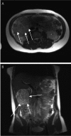
Metanephric adenoma of the kidney: an unusual diagnostic challenge
- PMID: 21139840
- PMCID: PMC2994510
- DOI: 10.4081/rt.2010.e38
Metanephric adenoma of the kidney: an unusual diagnostic challenge
Abstract
Although metanephric adenoma (MA) is a rare, benign neoplasm of epithelial cells, it is often difficult to distinguish this entity from other malignant neoplasms preoperatively. We report a case of a large renal mass for which preoperative diagnosis was indeterminate, with the differential diagnosis including Wilm's tumor, MA, and papillary renal cell carcinoma (PRCC). Accurate postoperative differentiation of MA from PRCC is critical because adjuvant therapy is considered after surgical resection of PRCC tumors.
Keywords: Wilm’s tumor; differential diagnosis.; metanephric adenoma; papillary renal cell carcinoma.
Conflict of interest statement
Conflict of interest: the authors report no conflicts of interest.
Figures


References
-
- Pins MR, Jones EC, Martul EV, et al. Metanephric adenoma-like tumors of the kidney: report of three malignancies with emphasis on discriminating features. Arch Pathol Lab Med. 1999;123:415–20. - PubMed
-
- Amin MB, Amin MB, Tamboli P, et al. Prognostic impact of histologic subtyping of adult renal epithelial neoplasms: an experience of 405 cases. Am J Surg Pathol. 2002;26:281–91. - PubMed
-
- Schmelz HU, Stoschek M, Schwerer M, et al. Metanephric adenoma of the kidney: case report and review of the literature. Int Urol Nephrol. 2005;37:213–7. - PubMed
-
- Renshaw AA, Freyer RD, Hammers AY. Metastatic metanephric adenoma in a child. Am J Surg Pathol. 2000;24:570–4. - PubMed
Publication types
LinkOut - more resources
Full Text Sources

