Primary breast lymphomas
- PMID: 21139885
- PMCID: PMC2994446
- DOI: 10.4081/rt.2009.e14
Primary breast lymphomas
Abstract
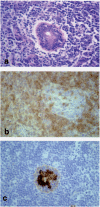
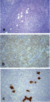
The diagnosis, prognostic factors, and optimal management of primary breast lymphomas (PBL) is difficult. Seven patients recorded at the Geneva Cancer Registry between 1973-1998 were reviewed. Five patient had diffuse large B-cell lymphoma, one a follicular lymphoma and one a MALT-lymphoma. All patients had clinical and radiological findings consistent with breast cancer and underwent mastectomy, which is not indicated in PBL. Diagnosis should be established prior to operative interventions, as fine needle aspiration missed the diagnosis for one patient and intra-operative frozen sections for 3 patients in our study. Five-year and 10-year overall survivals were 57% and 15%, respectively. Of the 3 patients who died from PBL, 2 had tumors that were Bcl-2 positive but Bcl-6 negative. All 3 surviving patients have positive Bcl-2 and Bcl-6 immunostaining, which could be important prognostic factors if confirmed by a larger study.
Keywords: bcl2.; bcl6; breast; cancer; lymphoma.
Figures


References
-
- Wiseman C, Liao KT. Primary lymphoma of the breast. Cancer. 1972;29:1705–12. - PubMed
-
- Bouchardy C. In: Cancer incidence in five continents. W S Parkin DM, Ferlay J, Raymond L, Young J, editors. Vol. VII. Lyon: International Agency for Research for Cancer; 1997. pp. 666–9.
-
- Oncology I.-O.I.C.O.D.F. 1st ed. Geneva: WHO; 1976.
-
- International Classification of Diseases R. ed. Geneva: WHO; 1967.
LinkOut - more resources
Full Text Sources

