

Case Reports
doi: 10.4081/rt.2009.e24.
Cystadenofibroma of the rete ovarii: a case report with review of literature
Affiliations
- PMID: 21139896
- PMCID: PMC2994433
- DOI: 10.4081/rt.2009.e24
Item in Clipboard
Case Reports
Cystadenofibroma of the rete ovarii: a case report with review of literature
Rare Tumors.
.
No abstract available
Keywords: cystadenofibroma; immunohistochemistry.; ovary; rete ovarii; rete testis.
Conflict of interest statement
Conflict of interest: the authors reported no potential conflict of interests.
Figures

Gross photograph showing bi-valved specimen depicting solid- cystic appearance.

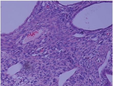
Microphotograph showing diffuse replacement of ovary by epithelial and stromal proliferation (H&E × 40).

Microphotograph showing variably sized irregular slit-like spaces lined by monolayered bland cuboidal epithelium and intervening mature fibrotic stroma (H&E
×200).

Microphotograph showing a markedly dilated cyst with papillae projecting into the cyst lumen (H&E ×200).

Microphotograph showing high power view of bland cuboidal lining of papillae (H&E ×400).

Microphotograph showing bland cuboidal lining of tubules with occasional ciliated cells (H&E ×600).
Microphotograph showing cellular fibrotic stroma, reminiscent of rete ovarii (H&E ×200).

Microphotograph showing focal smooth muscle fibers (highlighted by SMA immunostaining) in the stroma (H&E ×200).

Microphotograph showing intense and diffuse ER positivity in epithelial cells (H & DAB × 200).

Microphotograph showing intense and diffuse CK7 positivity in epithelial cells (H & DAB ×200).

Microphotograph showing no staining in epithelial cells for thrombomodulin (right), whereas the normal ovarian surface epithelial lining (left) is positive for it, acting as an
internal control (H & E ×200).

Microphotograph showing lack of a distinct basement membrane, highlighted by negative staining for laminin (H & DAB ×400).
References
-
- Rutgers JL, Scully RE. Cysts (cystadenomas) and tumours of rete ovarii. Int J Gynec Pathol. 1988;7:330–42. - PubMed
-
- Long GG. Apparent mesonephric duct (rete anlage) origin for cysts and proliferative epithelial lesions in the mouse ovary. Toxic Pathol. 2002;3:592–8. - PubMed
-
- Heatley MK. Adenomatous hyperplasia of the rete ovarii. Histopathol. 2000;36:383–4. - PubMed
-
- Jones EC, Murray SK, Young RH. Cysts and epithelial proliferations of the testicular collecting system (including rete testis) Semin Diagn Pathol. 2000;17:270–93. - PubMed
-
- Devouassoux-Shisheboran M, Silver SA, Tavassoli FA. Wolffian Adnexal Tumor, so called female adnexal tumour of probable Wolffian origin (FATWO): immunohistochemical evidence in support of a Wolffian origin. Hum Pathol. 1999;30:856–63. - PubMed
Publication types
LinkOut - more resources
Full Text Sources
