

Diagnosis of vulvar lesions by non-invasive optical analysis: a pilot study
- PMID: 21139902
- PMCID: PMC2994441
- DOI: 10.4081/rt.2009.e8
Diagnosis of vulvar lesions by non-invasive optical analysis: a pilot study
Abstract
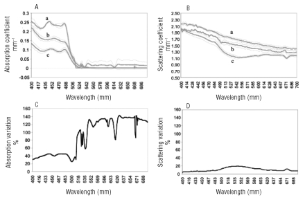
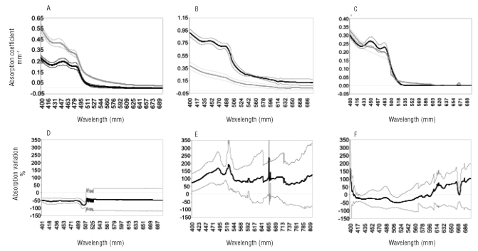
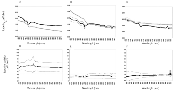
A procedure that could allow an early in vivo and non-invasive detection of vulvar lesions would be extremely useful. We tested an innovative optical method (Optiprobe), which uses a harmless, visible light source for the in vivo, on-line detection of minimal alterations in the structure of vulvar epithelium. A group of 3 female volunteers without gynecological symptoms were first screened to evaluate optical properties of normal vulvar tissue. Next, a group of 16 patients undergoing gynecological examination for vulvar lesions was evaluated by the Optiprobe at suspected sites before these sites were biopsied for histological analysis. Adjacent, non-involved sites were also measured to provide internal controls. Histological analysis of the biopsies identified one case that did not show obvious alterations, 4 cases of high-grade vulvar intraepithelial neoplasia (VIN), 5 cases of vulvitis, and 6 cases of lichen sclerosis (LS).The optical properties of the VIN cases were significantly different from those of controls, due to a decrease in the absorption spectra and an increase in the scattering spectra. In contrast, a significant increase in the absorption spectra and a decrease in the scattering spectra were observed in the cases of vulvitis. In the LS cases, the absorption spectra were as in controls, whereas the scattering spectra were significantly decreased. We conclude that the Optiprobe provides a useful tool for a rapid and non-invasive detection of vulvar alterations. The method should contribute to reduce the number of biopsies and to facilitate the long-term follow-up of vulvar lesions.
Keywords: absorption spectra; non invasive diagnosis; optical analysis; scattering spectra.; vulvar intraepithelial neoplasia.
Figures
Similar articles
-
Histopathologic study of thin vulvar squamous cell carcinomas and associated cutaneous lesions: a correlative study of 48 tumors in 44 patients with analysis of adjacent vulvar intraepithelial neoplasia types and lichen sclerosus.Am J Surg Pathol. 2006 Mar;30(3):310-8. doi: 10.1097/01.pas.0000180444.71775.1a. Am J Surg Pathol. 2006. PMID: 16538050
-
In the absence of (early) invasive carcinoma, vulvar intraepithelial neoplasia associated with lichen sclerosus is mainly of undifferentiated type: new insights in histology and aetiology.J Clin Pathol. 2007 May;60(5):504-9. doi: 10.1136/jcp.2005.031989. Epub 2006 May 19. J Clin Pathol. 2007. PMID: 16714399 Free PMC article.
-
Vulvar lichen sclerosus and squamous cell carcinoma: a cohort, case control, and investigational study with historical perspective; implications for chronic inflammation and sclerosis in the development of neoplasia.Hum Pathol. 1998 Sep;29(9):932-48. doi: 10.1016/s0046-8177(98)90198-8. Hum Pathol. 1998. PMID: 9744309 Review.
-
Vulvar intraepithelial neoplasia of the simplex (differentiated) type: a clinicopathologic study including analysis of HPV and p53 expression.Am J Surg Pathol. 2000 Mar;24(3):429-41. doi: 10.1097/00000478-200003000-00013. Am J Surg Pathol. 2000. PMID: 10716158
-
[Review of precancerous vulvar lesions].Cesk Patol. 2012 Jan;48(1):15-21. Cesk Patol. 2012. PMID: 22716003 Review. Czech.
References
-
- Eifel P, Levenback C, editors. Cancer of the lower genital tract. B.D.Inc; London Hamilton: 2001. pp. 1–8.
-
- Silverberg E. Statistical and epidemiological information on gynecological cancer. American Cancer Society. 1986;80:9–9.
-
- Cramer D. Epidemiology of the gynaecologic cancers. Com Theor. 1978;4:9–17. - PubMed
-
- Hart WR. Vulvar intraepithelial neoplasia: historical aspects and current status. Int J Gynecol Pathol. 2001;20:16–30. - PubMed
-
- Joura EA. Epidemiology, diagnosis and treatment of vulvar intraepithelial neoplasia. Curr Opin Obstet Gynecol. 2002;14:39–4. - PubMed
LinkOut - more resources
Full Text Sources
