

Case Reports
doi: 10.4081/rt.2009.e9.
Congenital giant melanocytic nevi
Affiliations
- PMID: 21139903
- PMCID: PMC2994429
- DOI: 10.4081/rt.2009.e9
Item in Clipboard
Case Reports
Congenital giant melanocytic nevi
Rare Tumors.
.
Abstract
Nevi are common skin tumors caused by abnormal overgrowth of cells from the epidermal and dermal layers of the skin. Most nevi are benign, but some pre-cancerous nevi must be monitored or removed. The giant congenital nevus is greater than 10 cm in size, pigmented and often hairy. Between 4% and 6% of these lesions will develop into a malignant melanoma. Since approximately 50% of the melanoma develop by the age of two, and 80% by the age of seven, early removal is recommended. The objective of this paper is to present a unique case of giant nevi and their surgical management.
Keywords: congenital.; giant; melanoma; nevus.
Figures
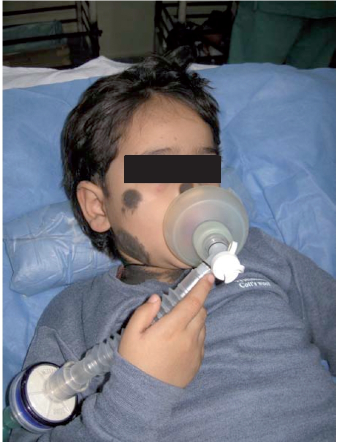
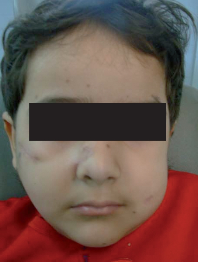
Pre-operative photograph of the patient on OT table.
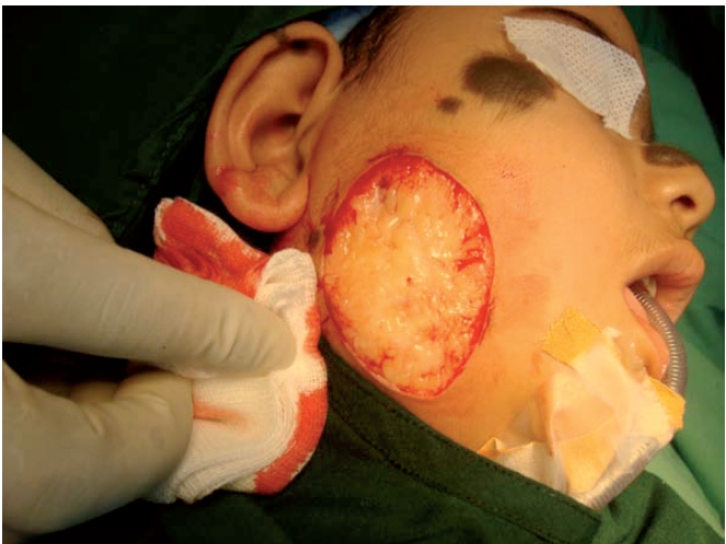
Intra-operative photograph of the same patient.
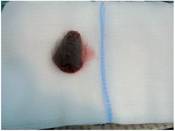
Resected specimen of the same patient.
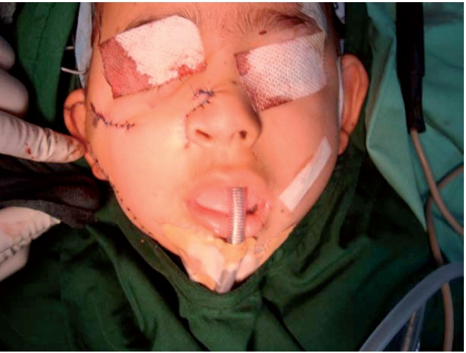
Post-operative photograph of the same patient on OT table.
Post-operative photograph of the same patient.
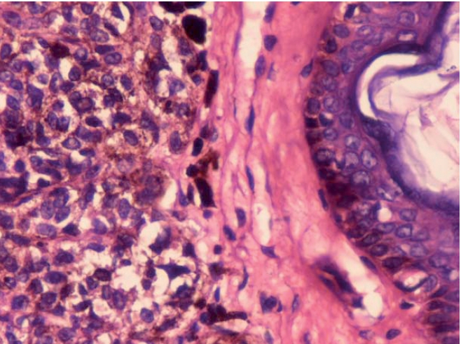
(40×) Histopathological examination shows nevi cells are separated with connective tissue layer.
Similar articles
-
Giant Congenital Melanocytic Nevi Successfully Treated with Combined Laser Therapy.Indian Dermatol Online J. 2020 Jan 13;11(1):79-82. doi: 10.4103/idoj.IDOJ_107_19. eCollection 2020 Jan-Feb. Indian Dermatol Online J. 2020. PMID: 32055515 Free PMC article.
-
Giant congenital melanocytic nevi: a case report.J Clin Diagn Res. 2013 Jan;7(1):154-5. doi: 10.7860/JCDR/2012/4832.2693. Epub 2012 Nov 1. J Clin Diagn Res. 2013. PMID: 23450701 Free PMC article.
-
Congenital melanocytic nevi: clinical and histopathologic features, risk of melanoma, and clinical management.J Am Acad Dermatol. 2005 Feb;52(2):197-203. doi: 10.1016/j.jaad.2004.07.020. J Am Acad Dermatol. 2005. PMID: 15692463 Review.
-
The controversial management of giant congenital melanocytic nevi. When would it be better "to wait and see"?G Ital Dermatol Venereol. 2013 Apr;148(2):203-7. G Ital Dermatol Venereol. 2013. PMID: 23588146
-
Significant melanocytic lesions in infancy, childhood, and adolescence.Dermatol Clin. 1986 Jan;4(1):29-44. Dermatol Clin. 1986. PMID: 3521978 Review.
Cited by
-
Reconstruction of a Giant Congenital Melanocytic Nevus Defect With a Submental Flap in a Global Health Setting.Cureus. 2021 Jul 30;13(7):e16751. doi: 10.7759/cureus.16751. eCollection 2021 Jul. Cureus. 2021. PMID: 34513374 Free PMC article.
-
Topical therapy for regression and melanoma prevention of congenital giant nevi.Cell. 2022 Jun 9;185(12):2071-2085.e12. doi: 10.1016/j.cell.2022.04.025. Epub 2022 May 12. Cell. 2022. PMID: 35561684 Free PMC article.
-
A large-scale collection of giant congenital melanocytic nevi: Clinical and histopathological characteristics.Exp Ther Med. 2020 Jan;19(1):313-318. doi: 10.3892/etm.2019.8198. Epub 2019 Nov 14. Exp Ther Med. 2020. PMID: 31853305 Free PMC article.
-
Giant congenital melanocytic nevus in a Cameroonian child: a case report.J Med Case Rep. 2018 Jun 23;12(1):175. doi: 10.1186/s13256-018-1707-y. J Med Case Rep. 2018. PMID: 29933750 Free PMC article.
References
-
- Chunj C, Forte AJ, Narayan D, et al. A review. J Craniofac Surg. 2006:1210–15. - PubMed
-
- Gosain AK, Santoro TD, Larson DL, et al. Congenital Nevi: a 20 - year experience and an algorithm for their management. Plast Reconstr Surg. 2001;108:622–36. - PubMed
-
- McWhorter HE, Woolner LB. Pigmented Nevi, Juvenile Melanoma and Malignant melanoma in children. Cancer. 2006;7:564–85. - PubMed
-
- Zemtsov A, Lorig R, Bergfeld W, et al. Magnetic resonance imaging of cutaneous melanocytic lesions. J Dermatol Surg Oncol. 1989;15:854–8. - PubMed
-
- Shaw M. Malignant melanoma arising from a giant hairy naevus. Br J Plast Surg. 1962;15:426–31. - PubMed
Publication types
LinkOut - more resources
Full Text Sources
