

Primary retroperitoneal mullerian adenocarcinoma
- PMID: 21139951
- PMCID: PMC2994487
- DOI: 10.4081/rt.2010.e6
Primary retroperitoneal mullerian adenocarcinoma
Abstract
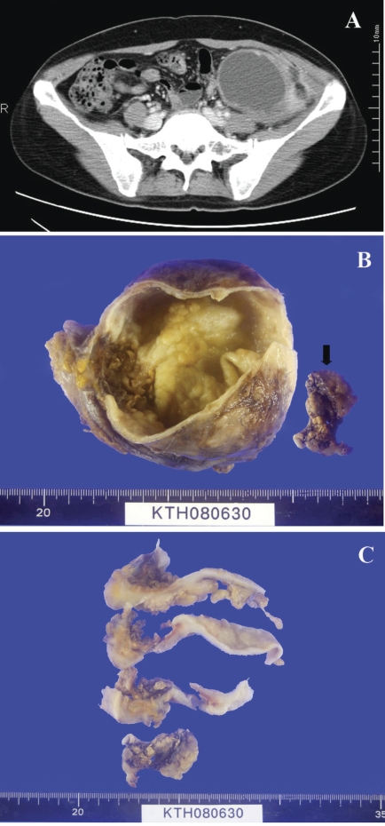
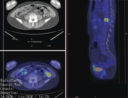
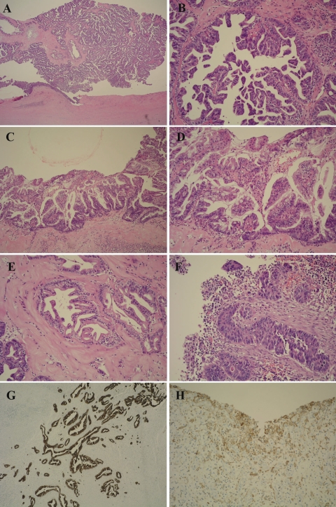
Mullerian tumors are extremely rare malignancies in the retroperitoneum. We report a case of a 46-year old woman who presented with an eight year history of lower abdominal mass. Ultrasonography (US) and computed tomography (CT) demonstrated a 15×10 cm cystic mass in the left lower retroperitoneum. As serial percutaneous needle aspiration cytology was negative for malignancy, she was observed for seven years. Eleven months ago, the mass was excised. The histopathology was reported as mucinous adenocarcinoma of the retroperitoneum. Six cycles of intraperitoneal (IP) chemotherapy was administered during the last six months after diagnosis of recurrence by aspiration cytology and high serum tumor markers (CEA, CA19-9). A few days ago, positron emission tomographic (PET) scanning showed evidence of local recurrence and single vertebral metastasis, so she was admitted again for systemic chemotherapy. Meticulous revision of additional sections of the tumor revealed papillary, serous, mucinous, and endometrioid subtypes of the mullerian adenocarcinoma. To our knowledge, there has been no similar case described in the literature.
Keywords: adenocarcinoma; mullerian tumor; retroperitoneum.
Figures
References
-
- Vaidya AP, Horowitz NS, Oliva E, et al. Uterine malignant mixed mullerian tumors should not be included in studies of endometrial carcinoma. Gynecol Oncol. 2006;103:684–7. - PubMed
-
- Neesham D, Kerdemelidis P, Scurry J. Primary malignant mixed Mullerian tumor of the vagina. Gynecol Oncol. 1998;70:303–7. - PubMed
-
- Clement PB, Zubovits JT, Young RH, Scully RE. Malignant mullerian mixed tumors of the uterine cervix: a report of nine cases of a neoplasm with morphology often different from its counterpart in the corpus. Int J Gynecol Pathol. 1998;17:211–22. - PubMed
-
- Gagner JP, Mittal K. Malignant mixed Mullerian tumor of the fimbriated end of the fallopian tube: origin as an intraepithelial carcinoma. Gynecol Oncol. 2005;97:219–22. - PubMed
-
- Mok JE, Kim YM, Jung MH, et al. Malignant mixed mullerian tumors of the ovary: experience with cytoreductive surgery and platinum-based combination chemotherapy. Int J Gynecol Cancer. 2006;16:101–5. - PubMed
Publication types
LinkOut - more resources
Full Text Sources
Research Materials
