Case Reports
. 2010 Sep 30;2(3):e48.
doi: 10.4081/rt.2010.e48.

Pediatric primary intramedullary spinal cord glioblastoma

Affiliations
Case Reports

Pediatric primary intramedullary spinal cord glioblastoma

Robert Lober et al. Rare Tumors. .

Abstract

Spinal cord tumors in pediatric patients are rare, representing less than 1% of all central nervous system tumors. Two cases of pediatric primary intramedullary spinal cord glioblastoma at ages 14 and 8 years are reported. Both patients presented with rapid onset paraparesis and quadraparesis. Magnetic resonance imaging in both showed heterogeneously enhancing solitary mass lesions localized to lower cervical and upper thoracic spinal cord parenchyma. Histopathologic diagnosis was glioblastoma. Case #1 had a small cell component (primitive neuroectodermal tumor-like areas), higher Ki67, and p53 labeling indices, and a relatively stable karyotype with only minimal single copy losses involving regions: Chr8;pter-30480019, Chr16;pter-29754532, Chr16;56160245-88668979, and Chr19;32848902-qter on retrospective comparative genomic hybridization using formalin-fixed, paraffin-embedded samples. Case #2 had relatively bland histomorphology and negligible p53 immunoreactivity. Both underwent multimodal therapy including gross total resection, postoperative radiation and chemotherapy. However, there was no significant improvement in neurological deficits, and overall survival in both cases was 14 months.This report highlights the broad histological spectrum and poor overall survival despite multi modality therapy. The finding of relatively unique genotypic abnormalities resembling pediatric embryonal tumors in one case may highlight the value of genome-wide profiling in development of effective therapy. The differences in management with intracranial and low-grade spinal cord gliomas and current management issues are discussed.

Keywords: glioblastoma; intramedullary; pediatric; spinal cord.


Figures
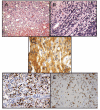
Figure 1
Figure 1
Case #1. (A) Densely cellular neoplasm, with hyperchromatic and pleomorphic astrocytic cells in a well-formed fibrillary background meshwork, with focal necrosis (H&E stain; 100× magnification). (B) Densely cellular infiltrating portion of the neoplasm, with markedly hyperchromatic and pleomorphic overlapping astrocytic cells in a rather sparse fibrillary background with focal necrosis, including small cells focally resembling a primitive neuroectodermal tumor but with microvascular proliferation (H&E stain; 200×). (C) The tumor shows diffuse glial fibrillary acidic protein (GFAP) positivity with dense fibrillary network in portions (GFAP stain; 400×). (D) The tumor shows a high Ki67 (MIB1) immunohistochemical nuclear labeling index of up to about 30% (Ki67 stain; 200×). (E) The tumor shows a high p53 immunohistochemical nuclear labeling index of up to about 70% (p53 stain; 200×).
Figure 2
Figure 2
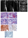
Case #2. (A) T1 weighted sagittal magnetic resonance images without contrast show an intramedullary hypointense mass lesion present within the central portion of the cervical spinal cord from C3 to T3 levels, producing expansion. (B) T1 weighted sagittal MR images with contrast show an intramedullary enhancing mass lesion present within the central portion of the cervical spinal cord, originating from the region of the central canal, producing expansion. The spinal cord measures 13 mm at the greatest AP diameter. The mass shows rim enhancement with central cystic changes and is surrounded by normal-appearing spinal cord. This mass extends from the level of the obex inferiorly to the T4 vertebral body. The maximum bulk of the mass is present from C3 to T3 levels. This mass produces remodeling of the posterior vertebral bodies most notably at C4 to C7. (C) T2 weighted sagittal MRI showing an expansile intramedullary mass lesion present within the cervical spinal cord. (D) Post-contrast T1 weighted axial MRI showing an enhancing expansile intramedullary mass lesion present within the central portion of the spinal cord. (E) Post-contrast T1 weighted axial MRI showing a ring-enhancing expansile intramedullary mass lesion present within the central portion of the spinal cord. (F) Moderate to focal densely cellular neoplasm with moderately hyperchromatic and pleomorphic loosely arranged astrocytic cells, with focal marked microvascular proliferation (H&E stain; 200×). (G) Areas of necrosis within the astrocytic neoplasm. No pseudopalisading is appreciated (H&E stain; 100×). (H) Moderate to focal densely cellular neoplasm with moderately hyperchromatic and pleomorphic astrocytic cells, with more than one mitosis in one high-power field, and with focal moderate microvascular proliferation (H&E stain; 400×). (J) Moderately hyperchromatic and pleomorphic astrocytic cells, with more than one mitosis in one high-power field (H&E stain; 400×). (K) Irregular infiltration of diffusely immunopositive neoplastic astrocytic cells, with negative images of hyperplastic vessels (GFAP stain; 100×). (L) The tumor shows a high Ki67 (MIB1) immunohistochemical nuclear labeling index of up to about 20% (Ki67 stain; 200×). (M) The tumor shows a negligible p53 immunohistochemical nuclear labeling index of less than 5% (p53 stain; 200×).

Similar articles

Cited by

References

    1. Epstein FJ. Philadelphia: W.B. Saunders; 1994. Pediatric Neurosurgery.
    1. Helseth A, Mørk SJ. Primary intraspinal neoplasms in Norway, 1955 to 1986. A population-based survey of 467 patients. J Neurosurg. 1989;71:842–5. - PubMed
    1. Merchant TE, Nguyen D, Thompson SJ, et al. High-grade pediatric spinal cord tumors. Pediatr Neurosurg. 1999;30:1–5. - PubMed
    1. Minehan KJ, Brown PD, Scheithauer BW, et al. Prognosis and treatment of spinal cord astrocytoma. Int J Radiat Oncol Biol Phys. 2009;73:727–33. - PubMed
    1. Tendulkar RD, Panandiker AS, Wu S, et al. Irradiation of pediatric high-grade spinal cord tumors. Int J Radiat Oncol Biol Phys 2010. [Epub ahead of print] - PMC - PubMed

Publication types