. 2010 Nov 4:16:2301-16.

HuB/C/D, nPTB, REST4, and miR-124 regulators of neuronal cell identity are also utilized in the lens

Affiliations

HuB/C/D, nPTB, REST4, and miR-124 regulators of neuronal cell identity are also utilized in the lens

Claudine L Bitel et al. Mol Vis. .

Abstract

Purpose: An interlocking network of transcription factors, RNA binding proteins, and miRNAs globally regulates gene expression and alternative splicing throughout development, and ensures the coordinated mutually exclusive expression of non-neural and neuronal forms of these factors during neurogenesis. Striking similarities between lens fiber cell and neuron cell morphology led us to determine if these factors are also used in the lens. HuR and polypyrimidine tract binding protein (PTB) have been described as 'global regulators' of RNA alternative splicing, stability, and translation in non-neuronal (including ectodermal) tissues examined to date in diverse species, and REST/NRSF (RE-1 Silencing Transcription Factor/Neuron Restrictive Silencing Factor) represses>2,000 neuronal genes in all non-neuronal tissues examined to date, but has not included the lens. During neurogenesis these factors are replaced by what has been considered neuron-specific HuB/C/D, nPTB, and alternatively spliced REST (REST4), which work with miR-124 to activate this battery of genes, comprehensively reprogram neuronal alternative splicing, and maintain their exclusive expression in post-mitotic neurons.

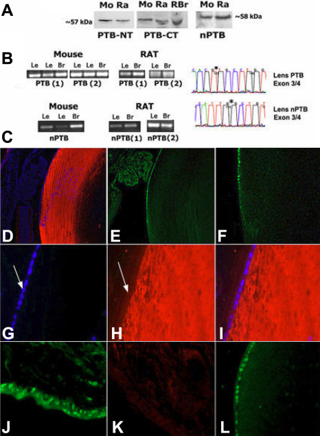
Methods: Immunoprecipitation, western blot, immunofluorescence, and immunohistochemistry were used to determine the expression and distribution of proteins in mouse and rat lenses. Mobility shift assays were used to examine lenses for REST/NRSF DNA binding activity, and RT-PCR, DNA sequencing, and northern blots were used to identify RNA expression and alternative splicing events in lenses from mouse, rat, and goldfish (N. crassa).

Results: We demonstrated that REST, HuR, and PTB proteins are expressed predominantly in epithelial cells in mouse and rat lenses, and showed these factors are also replaced by the predominant expression of REST4, HuB/C/D and nPTB in post-mitotic fiber cells, together with miR-124 expression in vertebrate lenses. REST-regulated gene products were found to be restricted to fiber cells where REST is decreased. These findings predicted nPTB- and HuB/C/D-dependent splicing reactions can also occur in lenses, and we showed Neuronal C-src and Type 1 Neurofibromatosis 1 splicing as well as calcitonin gene related peptide (CGRP) and neural cell adhesion molecule (NCAM-180) alternative transcripts in lenses. Transgenic mice with increased HuD in lens also showed increased growth associated protein 43 (GAP43) and Ca++/Calmodulin dependent kinase IIα (CamKIIα) HuD target gene expression in the lens, similar to brain.

Conclusions: The present study provides the first evidence this fundamental set of regulatory factors, previously considered to have a unique role in governing neurogenesis are also used in the lens, and raises questions about the origins of these developmental factors and mechanisms in lens and neuronal cells that also have a basic role in determining the neuronal phenotype.


Figures

Figure 1
Figure 1
Structure of the eye and cellular lens; organization of vertebrate lenses. Small cuboidal epithelial cells cover the anterior surface. At the anterior/posterior equator, these cells exit the cell cycle and begin to elongate as they move into the interior. A few hundred microns into the lens, fiber cells undergo a final stage of terminal differentiation where they lose cell nuclei and organelles. At right is a histological section of an adult mouse eye.
Figure 2
Figure 2
REST (NRSF) regulated neuronal genes are activated in the lens. A: Immunoblot detection of Synapsin 1 in lens and brain (a: Millipore; b: BD Biosciences). B: Amplification of Synapsin 1 transcripts from lens and brain. C: Representative sequence identifying exon junction in cDNA products. D: Immunoblot detection of neuronal βIII-tubulin in lens, and brain (a: rabbit antibody, Sigma; b: chicken antibody, Aves). E: Amplification of neuronal βIII-tubulin transcripts from lens and brain. F: Region from complete amplified product sequence identifying in frame exon junction in amplified transcripts. G: Amplification of Synaptotagmin 1 and Synaptophysin 1 transcripts from lens and brain. H: Region from amplified product sequence identifying in frame exon junctions in amplified transcripts. Asterisks indicate splice junctions.
Figure 3
Figure 3
REST regulated neuronal genes are activated in post-mitotic fiber cells. A: Hematoxylin and eosin stained mouse lens. B: Mouse mAb βIII-tubulin. C: Rabbit mAb anti-βIII-tubulin. D: Chicken anti-βIII-tubulin. E: Anti-Synaptophysin 1. F: Mouse mAb Synaptotagmin 1. G: Rabbit anti-Synapsin 1. H: DAPI as in G. I: No primary antibody control (lo), DAPI. J: Synapsin 1 in retinal neuronal layers (autofluorescence is seen in photoreceptor layer). K: mAb βIII-tubulin. L: Synaptotagmin 1 in peripheral fiber cells.
Figure 4
Figure 4
REST/NRSF, and neuron-specific alternatively-spliced REST4 are produced in lenses; identification of functional NRSE binding activity in the lens. A: Immunoblot and IP detection of REST and REST4 in the lens. Left panels: mAb anti-REST and anti-REST4 3121. Right panels: Immunoblot of lens proteins immunoprecipitated with NH2-terminal anti-REST mAb, probed with mAb anti-REST or anti-REST4 3122. B: Detection of REST in lenses using anti-COOH-terminus REST. C: Amplified REST4 (R4A, R4B) and REST (RA) transcripts from lenses. D: Gel mobility shift assay identifying REST:NRSE DNA binding activity in lens extracts. NRSE and nucleotide-substituted NRSE sequences are shown below. Asterisk indicates mobility shifted complexes. NRSE and nucleotide substituted competitor were added at 25- and 100-fold excess.
Figure 5
Figure 5
Mutually exclusive expression of REST in progenitor epithelial cells, and alternatively spliced REST4 in post-mitotic elongating lens fiber cells. A, B: Anti-C-terminal REST (100×); nuclei are stained with DAPI in panel A. C: Anti-REST4 3121 (50×). D: Anti-COOH-terminal REST (200×). E: Anti-REST4 3122 (200×). F: REST detected in rat skin. G: REST detected in Cornea. H: Overlapping REST4 and tubulin detection in lens fiber cells; cell nuclei are DAPI stained. I: HuR detected in rat skin. J: Anti-REST. K: Anti-REST4; nuclei are stained with DAPI in lens.
Figure 6
Figure 6
PTB (PTBP1) is expressed in progenitor epithelial cell and neuronal nPTB (PTBP2) in post-mitotic lens fiber cells. A: Immunoblot detection of PTB and nPTB in mouse and rat lens and brain tissue. PTB-NT: anti-NH2-terminus PTB, PTB-CT: anti-COOH-terminus PTB, and anti-nPTB. B: Left: Amplified PTB and nPTB transcripts from lens and brain. Right: Region from amplified DNA sequence product identifying in frame exon junctions in lens. C: Immunofluorescence detection of PTB and nPTB in the lens. D: anti-nPTB (100×), E: anti-PTB-NT (100×), F: anti-PTB-CT (100×), G: DAPI nuclear stain/no 1o control (200×), H: anti-nPTB (200×), I: overlay panel G and H. J: For comparison PTB is detected in cell nuclei in rat skin, K: anti-nPTB detects little or no protein in rat skin, L: Detection of PTB in cell nuclei in epithelial cells on the anterior lens surface.
Figure 7
Figure 7
HuR is expressed in progenitor lens epithelial cells, and neuronal HuB/C/D in post-mitotic elongating fiber cells. A: Amplification of HuB, HuC, and HuD transcripts from lens and brain; right: Region from sequenced product showing in frame exon junctions in amplified transcripts. B: Amplification of the HuD target transcripts: GAP43 and CamKIIα from wt mouse and rat lens and brain. C: Immunoblot detection of HuR in lens and brain. D: Immunoblot of HuB/C/D in lens and brain (Human anti-HuB/C/D). E: GAP43 protein detected in wt lens and brain. F: Increased expression of GAP43 and CamKIIα detected on immunoblots of wt versus transgenic mouse lenses expressing myc-tagged HuD in the lens; unchanged GAPDH levels are shown for comparison. G: Immunofluorescence detection in rat lens: H: mAb anti-HuR (200×), I: DAPI stained nuclei as in panel H (100×), J: Human anti-HuB/C/D, K: DAPI stained nuclei as in panel J (200×), L: Human anti-HuB/C/D (100×), M: anti-COOH-terminal REST, N: overlay of panels L and M, O: Syn1 expression in post-mitotic fiber cells (BD Biosciences, 200×), P: GAP43 in wt lens, Q: DAPI stained nuclei as in panel P, R: CamKIIα in wt lens, S: mAb anti-HuD detection in wt mouse lens (L-P, Santa Cruz).
Figure 8
Figure 8
HuB/C/D and nPTB dependent alternative splicing also occurs in mouse and rat lens. A: Neuronal alternative splicing of Nf1 Type 1 transcripts omits a 63 bp exon. Type 2 transcripts include this 63 bp exon, B: Amplification of Nf1 transcripts from lens and brain with primers corresponding to exons adjacent to the insert exon, C: Sequence of amplified Nf1 product from lens identifying Type 1 splicing, and omission of the 63 bp exon. D: In neurons, alternative splicing skips exon 4 to produce CGRP transcripts, E: Amplification of CGRP transcripts from lens and brain, F: Sequences identifying alternatively spliced 3–5 exon junctions in CGRP transcripts amplified from lenses. Alternative splicing of Neuronal C-src includes18 bp N1 exon. G: Amplification of C-src transcripts from lens and brain with primers corresponding to adjacent exons, H: DNA sequence of amplified Neuronal C-src product identifying the N1 exon in the isoform produced in lenses. I: nPTB-dependent alternative splicing of NCAM-180 transcripts includes an 801 bp exon, J: Amplified NCAM-180 transcripts from lens and brain, K: Sequence of amplified NCAM-180 transcripts from lens identifying the 5′ splice junction that includes the 801 bp exon, L: Immunoblot of NCAM-180 protein in rat lens using mAb anti-NCAM-180 specific antibody.
Figure 9
Figure 9
miR-124 in vertebrate lenses detected on northern blots. A: Left: Total RNA from N. crassa (goldfish) lenses resolved on acrylamide gels. Highly represented RNAs are stained with ethidium bromide indicated by arrowhead. Right: miR-124 detected with labeled probe. B: Left: Ethidium bromide stained RNAs from rat lens and brain. Right: miR-124 detected with radiolabeled miR-124 probe. Lower asterisk: ~22 bp nucleotide miR-124. Upper asterisk: ~76 bp precursor in rat brain. Probes, and negative controls showing no lens expression of muscle-specific miR-1, are described elsewhere [39].
Figure 10
Figure 10
Model of regulatory interactions between factors expressed in neural progenitors and lens epithelial cells versus post-mitotic neurons and lens fiber cells. In neurons REST/NRSF transcription factors, HuR-HuB/C/D and PTB-nPTB RNA binding proteins, with miR-124 form a network that differentially regulates non-neural and neuron-specific alternative splicing and gene expression. REST suppresses >1,500 neuronal genes in non-neuronal cells throughout the body. In post-mitotic neurons REST decreases, and neuron-specific alternatively spliced REST4 is produced, further relieving repression of these genes. Ubiquitous HuR and PTB that promote non-neural splicing are also replaced by neuron-specific HuB/C/D and nPTB in post-mitotic neurons, which leads to a comprehensive reprogramming of neuron-specific alternative splicing. In non-neural cells, PTB alters nPTB transcript splicing to tag them for nonsense mediated decay. In post-mitotic neurons, REST repression of miR-124 expression is alleviated, allowing miR-124 to suppress hundreds of non-neuronal transcripts, including PTB.

Similar articles

Cited by

References

    1. Alberts B. Molecular Biology of the Cell. Fourth ed. London: Garland Science; 2007.
    1. Boutz PL, Stoilov P, Li Q, Lin CH, Chawla G, Ostrow K, Shiue L, Ares M, Jr, Black DL. A post-transcriptional regulatory switch in polypyrimidine tract-binding proteins reprograms alternative splicing in developing neurons. Genes Dev. 2007;21:1636–52. - PMC - PubMed
    1. Chen ZF, Paquette AJ, Anderson DJ. NRSF/REST is required in vivo for repression of multiple neuronal target genes during embryogenesis. Nat Genet. 1998;20:136–42. - PubMed
    1. Fukao A, Sasano Y, Imataka H, Inoue K, Sakamoto H, Sonenberg N, Thoma C, Fujiwara T. The ELAV protein HuD stimulates cap-dependent translation in a Poly(A)- and eIF4A-dependent manner. Mol Cell. 2009;36:1007–17. - PubMed
    1. Jones FS, Meech R. Knockout of REST/NRSF shows that the protein is a potent repressor of neuronally expressed genes in non-neural tissues. Bioessays. 1999;21:372–6. - PubMed

Publication types

MeSH terms

LinkOut - more resources