. 2010 Nov 9:16:2328-41.

Gap junctions are selectively associated with interlocking ball-and-sockets but not protrusions in the lens

Affiliations

Gap junctions are selectively associated with interlocking ball-and-sockets but not protrusions in the lens

Sondip K Biswas et al. Mol Vis. .

Abstract

Purpose: Ball-and-sockets and protrusions are specialized interlocking membrane domains between lens fibers of all species studied. Ball-and-sockets and protrusions are similar in their shape, size, and surface morphology, and are traditionally believed to play a key role in maintaining fiber-to-fiber stability. Here, we evaluate the hypothesis that ball-and-sockets and protrusions possess important structural and functional differences during fiber cell differentiation and maturation.

Methods: Intact lenses of leghorn chickens (E7 days to P62 weeks old) and rhesus monkeys (1.5-20 years old) were studied with SEM, freeze-fracture TEM, freeze-fracture immunogold labeling (FRIL), and filipin cytochemistry for membrane cholesterol detection.

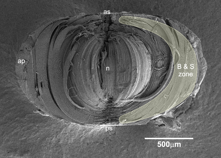
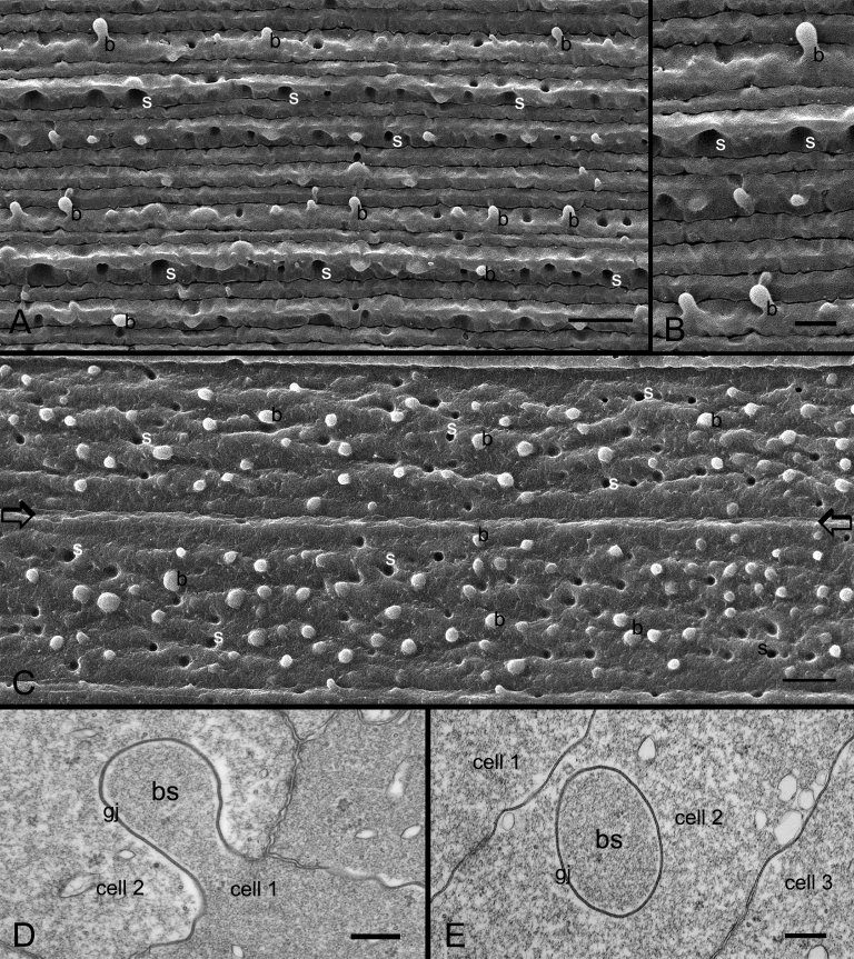
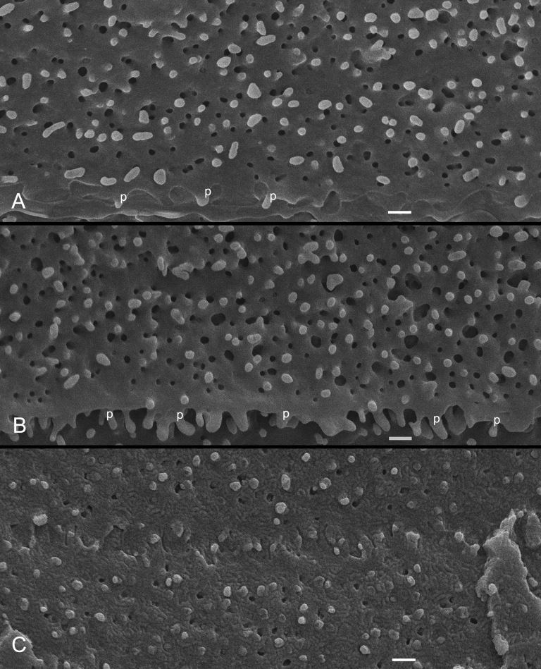
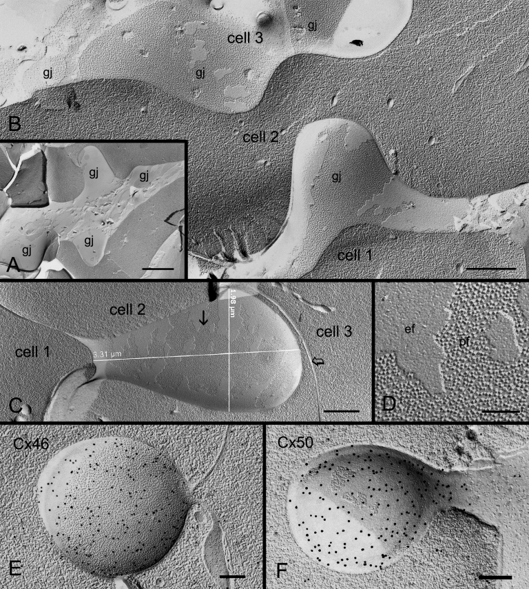
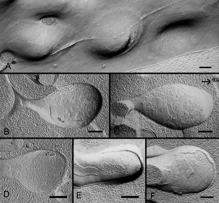
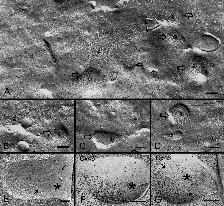
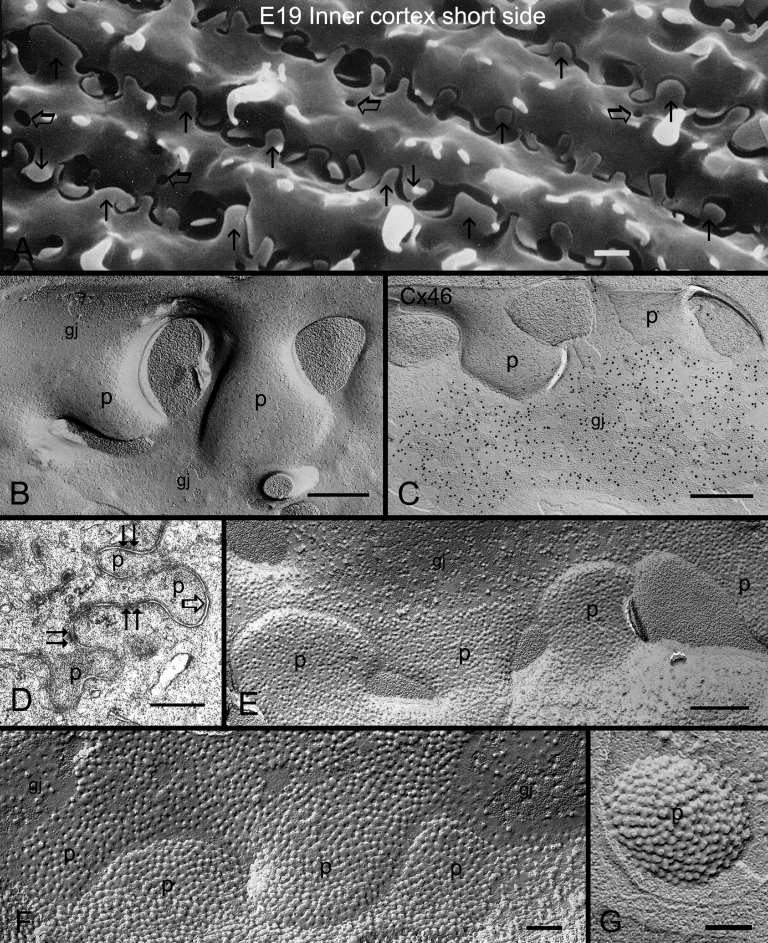
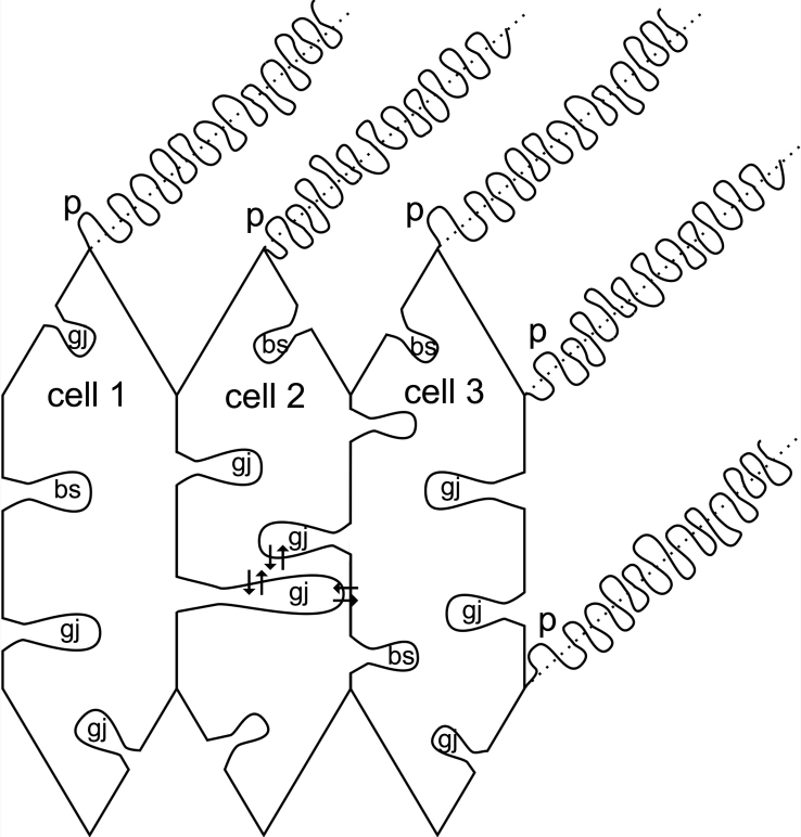
Results: SEM showed that ball-and-sockets were distributed along the long and short sides of hexagonal fiber cells, whereas protrusions were located along the cell corners, from superficial to deep cortical regions in both chicken and monkey lenses. Importantly, by freeze-fracture TEM, we discovered the selective association of gap junctions with all ball-and-sockets examined, but not with protrusions, in both species. In the embryonic chicken lens (E18), the abundant distribution of ball-and-socket gap junctions was regularly found in an approximate zone extending at least 300 μm deep from the equatorial surface of the superficial cortical fibers. Many ball-and-socket gap junctions often protruded deeply into neighboring cells. However, in the mature fibers of monkey lenses, several ball-and-sockets exhibited only partial occupancy of gap junctions with disorganized connexons, possibly due to degradation of gap junctions during fiber maturation and aging. FRIL analysis confirmed that both connexin46 (Cx46) and connexin50 (Cx50) antibodies specifically labeled ball-and-socket gap junctions, but not protrusions. Furthermore, filipin cytochemistry revealed that the ball-and-socket gap junctions contained different amounts of cholesterol (i.e., cholesterol-rich versus cholesterol-free) as seen with the filipin-cholesterol-complexes (FCC) in different cortical regions during maturation. In contrast, the protrusions contained consistently high cholesterol amounts (i.e., 402 FCCs/μm2 membrane) which were approximately two times greater than that of the cholesterol-rich gap junctions (i.e., 188 FCCs/μm2 membrane) found in ball-and-sockets.

Conclusions: Gap junctions are regularly associated with all ball-and-sockets examined in metabolically active young cortical fibers, but not with protrusions, in both chicken and monkey lenses. Since these unique gap junctions often protrude deeply into neighboring cells to increase membrane surface areas, they may significantly facilitate cell-to-cell communication between young cortical fiber cells. In particular, the large number of ball-and-socket gap junctions found near the equatorial region may effectively facilitate the flow of outward current toward the equatorial surface for internal circulation of ions in the lens. In contrast, a consistent distribution of high concentrations of cholesterol in protrusions would make the protrusion membrane less deformable and would be more suitable for maintaining fiber-to-fiber stability during visual accommodation. Thus, the ball-and-sockets and protrusions are two structurally and functionally distinct membrane domains in the lens.


Figures

Figure 1
Figure 1
SEM overview of fiber cell membrane surfaces of fractured intact embryonic chicken lens (E18) along the anterior/posterior (median sagittal) axis. This fractured axis is for examination of ball-and-sockets on the short sides of cortical fiber cells. An approximate zone for the abundant distribution of ball-and-sockets is outlined. In the equatorial region, the ball-and-socket zone extends at least 300 μm deep from the surface. In the images, B&S zone, ball-and-socket zone; AP, annular pad; N, nucleus; AS, anterior surface; PS, posterior surface.
Figure 2
Figure 2
SEM and thin-section TEM of ball-and-sockets in the embryonic and adult chicken lenses. A: In the embryonic chicken lens (E18), a representative scanning electron micrograph showing many ball-and-sockets distributed on the short sides of superficial cortical fibers at the equatorial region (approximately 100 μm from the surface). B: At higher magnification, the height and shape of the ball-and-sockets can be readily visualized from side-view. C: In the adult chicken lens (P42 weeks), numerous ball-and-sockets are seen distributed in rows on the two long sides of superficial cortical fibers at the equatorial region (approximately 200 μm from the surface). It is estimated that the number of ball-and-sockets (approximately 13 ball-and-sockets per 100 μm2 membrane) is not significantly different at the apical, equatorial and posterior regions along the anterior-posterior axis of any given superficial fiber cell. The border between two long-side fiber cells is marked by two arrows. Note that the height and the shape of these top-viewed ball-and-sockets cannot be readily appreciated as compared with those seen from the short sides in A and B. D: Thin-section TEM shows a ball-and-socket with the head and neck portions protruding into an adjacent cell. This ball-and-socket is completely occupied by a gap junction (gj). E: A cross-section of the ball-and-socket gap junction showing a considerable membrane extension of this junction into a narrow fiber cell. In the images, b is the ball, and s is the socket. The scale bar indicates 5 μm in A, C, 2 μm in B, and 200 nm in D and E.
Figure 3
Figure 3
SEM of ball-and-sockets in different cortical regions of monkey lens (20 year old). A: Superficial cortical fibers (approximately 100 μm from the surface), numerous ball-and-sockets are distributed on the long side of fiber cells. B: Intermediate cortical fibers (approximately 300 μm from the surface), a large number of ball-and-sockets are found on the long side of fiber cells. In this region, many protrusions (p) are also distributed along the corners of cortical fiber cells. C: However, in the deeper cortex (approximately 500 μm from the surface), ball-and-sockets display smaller number and size with degenerating appearance. The scale bars indicate 1 μm.
Figure 4
Figure 4
Freeze-fracture TEM and freeze-fracture immunogold labeling of Cx46 and Cx50 in ball-and-socket gap junctions of embryonic chicken lens fibers. A: Freeze-fracture TEM showing distribution of a cluster of ball-and-sockets containing gap junctions (gj). B: Higher magnification reveals the presence of gap junction plaque in the entire ball-and-socket domain (from cell 1) which protrudes into the cytoplasm of cell 2. The tip of this gap junction is in close proximity to the cell membrane of cell 3 which contains several flat gap junctions (gj). C: An elongated ball-and-socket gap junction, ~3.5 μm long and ~1.8 μm wide, from cell 1 protrudes deeply into the neighboring cell 2, and almost makes direct contact with the lateral cell membrane (open arrow) of cell 3. D: High magnification from the area marked by arrow in C showing gap junction particles (connexons) clearly seen on the P-face (pf) of the junctional membrane. E and F: FRIL shows the specific labeling of Cx46 and Cx50 antibodies in the ball-and-socket gap junctions, respectively. The scale bar indicates 1 μm in A; 500 nm in B and C; 100 nm in D, and 200 nm in E and F.
Figure 5
Figure 5
Freeze-fracture TEM and cholesterol distribution of ball-and-socket gap junctions in embryonic and adult chicken lens fibers. A: Ball-and-socket gap junction with loosely-packed configuration of connexons found in the outer cortex (0–400 μm from the surface). B: Ball-and-socket gap junction with a mixture of loosely-packed (open arrows) and crystalline-arranged (arrows) connexons found in the deeper region of the outer cortex. C: Ball-and-socket gap junction with crystalline-packed connexons (arrow) found in the inner cortex (400–800 μm). D: Cholesterol-rich ball-and-socket gap junction found in the outer cortex as determined by filipin cytochemistry in conjunction with freeze-fracture TEM. This gap junction exhibits loosely-packed connexons. E: Cholesterol-intermediate ball-and-socket gap junction found in the deeper region of the outer cortex. This gap junction displays distinct rows of crystalline-packed connexons (arrows) and few loose connexons. F and G: Cholesterol-free ball-and-socket gap junctions distributed in the inner cortex. These gap junctions contain the crystalline-packed configuration of connexons clearly seen on the P-face (pf) of the membrane in (F) and on the E-face (ef) of the membrane in (G). H and I: The presence of both cholesterol-rich and cholesterol-poor gap junctions in ball-and-sockets of the embryonic lens at E20. Several filipin-cholesterol-complexes (arrows) are indicated in the ball-and-socket in (I). The scale bars indicate 200 nm.
Figure 6
Figure 6
Freeze-fracture TEM of ball-and-socket gap junctions in monkey lens fibers. A: A cluster of three shallow ball-and-socket gap junctions are seen in superficial fibers of a young monkey (1.5 years old). B and C: Elongated ball-and-socket gap junctions with loosely-packed connexons in superficial cortical fibers of a mature monkey (20 years old). Arrow indicates the lateral cell membrane of adjacent cell. D, E, and F: Three different arrangements of connexons are associated with ball-and-sockets in the deeper mature cortical fibers: (D) A ball-and-socket completely occupied by connexons, (E) A ball-and-socket partially occupied by connexons, and (F) A ball-and-socket occupied by fragmentary gap junction plaques with disorganized connexons. Ball-and-socket gap junctions in E and F may be in a degradation stage. The scale bars indicate 200 nm.
Figure 7
Figure 7
Formation of ball-and-socket gap junctions in embryonic chicken lens fibers. A: An overview of superficial cortical fiber cells during early-stage formation of ball-and-socket gap junctions (gj) as seen in small invaginations and concavities (open arrows). Some small concavities (arrows) are devoid of connexons. Others contain loosely scattered connexons which are directly associated with nearby pools of flat membrane connexons, suggesting that ball-and-socket connexons perhaps migrate from nearby existing gap junctions of the flat membrane. B, C, and D: A representative profile showing examples of early ball-and-socket gap junction formation. Note that differing amounts of connexons are found distributed inside the concavities (arrow and open arrows), suggesting that concavities are formed before the migration (or insertion) of connexons. E: A well formed ball-and-socket domain is almost completely occupied by gap junction connexons. The non-junctional portion is indicated by asterisk. F and G: FRIL shows some immunogold particles for specific labeling of Cx46 antibody are scattered in the non-junctional portion (asterisk) inside the ball-and-socket, suggesting the presence of individual connexons for completion of gap junction formation in this area. The scale bars indicate 200 nm.
Figure 8
Figure 8
Structure and cholesterol distribution of protrusions in mature cortical fibers of the embryonic chicken lens fibers. A: SEM showing the distribution of numerous protrusions (arrows) from the corners of fiber cells in the inner cortex. Several small sockets (open arrows) of ball-and-sockets are also seen in the inner cortex. Note that two different sizes of protrusions from adjacent cells are often paired together for interlocking. B: Freeze-fracture TEM showing the absence of any gap junction on protrusions (p), although two gap junctions (gj) are found in close proximity to the protrusions (p). C: Freeze-fracture immunogold labeling confirms the absence of labeling for the Cx46 antibody in the protrusions (p). Instead, Cx46 antibody specifically labels the nearby large gap junction (gj). D: Thin-section TEM reveals the complex configuration of protrusions (p) without the association of gap junction (open arrow) with the protrusions. Surprisingly, several adherens junctions (paired arrows) are found associated with the neck portion of these protrusions. E: Filipin cytochemistry in conjunction with freeze-fracture TEM shows that a cluster of the protrusions (p) contain consistently high amounts of cholesterol (filipin-cholesterol-complexes, FCCs). F: At higher magnification, the protrusions display a high density of membrane cholesterol (i.e., 402 FCCs/μm2 membrane). Note that the adjacent gap junction (gj), classified as the cholesterol-rich subtype, contains only one half of FCCs (i.e., 188 FCCs/μm2 membrane) distributed in the protrusions. G: A top-viewed protrusion (p) in the cytoplasm showing a high density distribution of filipin-cholesterol-complexes. The scale bar indicates 1 μm in A, 500 nm in B, C, D, E, and 200 nm in F, and G.
Figure 9
Figure 9
A summary diagram depicting the important structural and functional differences between the ball-and-socket (BS) and protrusions (P) in hexagonal cortical fiber cells. Ball-and-sockets are distributed on both the long and short sides of fiber cells and are more frequent in the superficial than in the deeper cortex. They are generally larger in size but smaller in number than the protrusions distributed primarily along the corners. Structurally, gap junctions (gj) are present in all ball-and-sockets examined, but not in protrusions. Many elongated ball-and-socket gap junctions protrude deeply into neighboring fiber cells. Also, while the ball-and-socket gap junctions contain significantly different amounts of cholesterol during fiber differentiation and maturation, all protrusions examined consistently contain high amounts of membrane cholesterol. The cholesterol ratio between protrusions and the cholesterol-rich gap junctions seen in ball-and-sockets is approximately 2:1. It is suggested that the unique structural configuration of ball-and-socket gap junctions may significantly facilitate cell-to-cell communication (arrows) between metabolically active young fiber cells in the superficial cortex. Also, the large number of ball-and-socket gap junctions found near the equatorial region may effectively facilitate the flow of outward current toward the equatorial surface for internal circulation of ions in the lens. The presence of high cholesterol content in protrusions would make these domain membranes less deformable, and would be more suitable for maintenance of fiber-to-fiber stability during visual accommodation.

Similar articles

Cited by

References

    1. Rae JL. The electrophysiology of the crystalline lens. Curr Top Eye Res. 1979;1:37–90. - PubMed
    1. Goodenough DA. Lens gap junctions: a structural hypothesis for nonregulated low-resistance intercellular pathways. Invest Ophthalmol Vis Sci. 1979;18:1104–22. - PubMed
    1. Mathias RT, Riquelme G, Rae JL. Cell to cell communication and pH in the frog lens. J Gen Physiol. 1991;98:1085–103. - PMC - PubMed
    1. Mathias RT, White TW, Gong X. Lens gap junctions in growth, differentiation, and homeostasis. Physiol Rev. 2010;90:179–206. - PMC - PubMed
    1. Leeson TS. Lens of the rat eye: an electron microscope and freeze-etch study. Exp Eye Res. 1971;11:78–82. - PubMed

Publication types

LinkOut - more resources