Comparative Study
. 2011 Jul;82(7):1007-17.
doi: 10.1902/jop.2010.100577. Epub 2010 Dec 13.

Gene expression dynamics during bone healing and osseointegration

Affiliations
Comparative Study

Gene expression dynamics during bone healing and osseointegration

Zhao Lin et al. J Periodontol. 2011 Jul.

Abstract

Background: Understanding the molecular features of bone repair and osseointegration may aid in the development of therapeutics to improve implant outcomes. The purpose of this investigation is to determine the gene expression dynamics during alveolar bone repair and implant osseointegration.

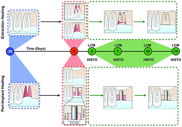
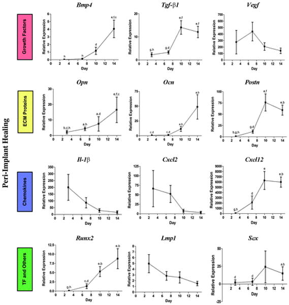
Methods: An implant osseointegration preclinical animal model was used whereby maxillary defects were created at the time of oral implant placement, while a tooth extraction socket healing model was established on the contralateral side of each animal. The surrounding tissues in the zone of the healing defects were harvested during regeneration for temporal evaluation using histology, immunohistochemistry, laser capture microdissection, and quantitative reverse transcription-polymerase chain reaction for the identification of a panel of 17 putative genes associated with wound repair.

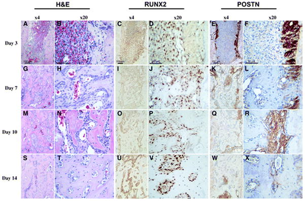
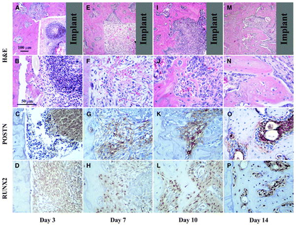
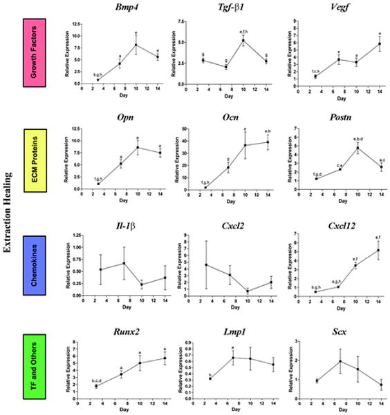
Results: In both models, three distinct expression patterns were displayed: 1) genes that are slowly increased during the healing process, such as bone morphogenetic protein 4, runt-related transcription factor 2, and osteocalcin; 2) genes that are upregulated at the early stage of healing and then downregulated at later stages, such as interleukin and chemokine (C-X-C motif) ligands 2 and 5; and 3) genes that are constitutively expressed over time, such as scleraxis. Although some similarities between osseointegration and tooth extraction socket were seen, distinct features developed and triggered a characteristic coordinated expression and orchestration of transcription factors, growth factors, extracellular matrix molecules, and chemokines.

Conclusions: Characterization of these events contributes to a better understanding of cooperative molecular dynamics in alveolar bone healing, and highlights potential pathways that could be further explored for the enhancement of osseous regenerative strategies.


Figures

Figure 1
Figure 1
Alveolar socket and peri-implant defect healing study models and timeline. The top and bottom panels represent the sequence of events that characterize the extraction and peri-implant healing models, respectively. The left first maxillary molar (M1) was extracted and allowed to completely heal for 28 days. On the healed ridge, an implant osteotomy was created that allowed implant placement and creation of a standardized peri-implant defect. In the contralateral side, M1 was extracted. LCM and histology (HISTO) methods were used for the analysis and evaluation of the healing area (black dotted line) at days 3, 7, 10, and 14.
Figure 2
Figure 2
Histologic evaluation of alveolar socket healing sites over time. Hematoxylin and eosin (H&E), RUNX2, and POSTN immunohistochemistry for tooth extraction site healing at 3, 7, 10, and 14 days (left panels, original magnification ×4; right panels, original magnification ×20). A and B) A clearly visible blood clot is noticeable at day 3. C and D) A significant number of RUNX2-positive cells are noticed within the alveolar socket populating the clot. Eand F) Remnants of the periodontal ligament can be clearly depicted by its strong POSTN staining at day 3. G through L) At day 7, the cell density in the defect area is higher and the POSTN- and RUNX2-positive cells start colocalizing within these areas. M through R) At day 10, the defect site seems to be filled by a condensed mesenchymal tissue. S through X) Finally, by day 14, an integration of the newly formed bone to the original socket walls is noticed.
Figure 3
Figure 3
Healing response for peri-implant repair sites at 3, 7, 10, and 14 days. A through D) Initially, inflammatory cells seem to dominate the defect area as depicted at day 3. E through H) By day 7, a loose fibrous connective tissue fills the defect and clear POSTN and RUNX2 staining is present. I through L) At day 10, RUNX2-positive cells are abundant and POSTN is gradually limited to the more immature tissue areas. M through P) Similar to the tooth extraction healing sites, at day 14, an integration of the newly formed bone to the walls of the defect is clear. Gray color in the top panels represents the implant location area (top panels, original magnification ×4; second, third, and fourth panels and rows, original magnification ×20).
Figure 4
Figure 4
Gene expression pattern of tooth extraction socket healing sites. ECM = extracellular matrix; TF = transcription factors. a = P <0.05 compared to day 3; b= P <0.05 compared to day 7; c = P <0.05 compared to day 10; d = P <0.05 compared to day 14; e = P <0.01 compared to day 3; f = P <0.01 compared to day 7; g = P <0.01 compared to day 10; h = P<0.01 compared to day 14.
Figure 5
Figure 5
Gene expression pattern of bone regenerative sites around implants. ECM = extracellular matrix; TF = transcription factors. a = P <0.05 compared to day 3; b = P <0.05 compared to day 7; c = P <0.05 compared to day 10; d = P <0.05 compared to day 14; e = P<0.01 compared to day 3; f = P <0.01 compared to day 7; g = P <0.01 compared to day 10; h = P <0.01 compared to day 14.

References

    1. Panetta NJ, Gupta DM, Longaker MT. Bone regeneration and repair. Curr Stem Cell Res Ther. 2010;5:122–128. - PubMed
    1. Gersbach CA, Phillips JE, García AJ. Genetic engineering for skeletal regenerative medicine. Annu Rev Biomed Eng. 2007;9:87–119. - PubMed
    1. Kimelman N, Pelled G, Helm GA, Huard J, Schwarz EM, Gazit D. Review: Gene- and stem cell-based therapeutics for bone regeneration and repair. Tissue Eng. 2007;13:1135–1150. - PubMed
    1. Secreto FJ, Hoeppner LH, Westendorf JJ. Wnt signaling during fracture repair. Curr Osteoporos Rep. 2009;7:64–69. - PMC - PubMed
    1. Lee J, Stavropoulos A, Susin C, Wikesjö UM. Periodontal regeneration: Focus on growth and differentiation factors. Dent Clin North Am. 2010;54:93–111. - PubMed

Publication types

MeSH terms