. 2010 Dec;5(10):1535-46.
doi: 10.2217/nnm.10.90.

Distribution and clearance of PEG-single-walled carbon nanotube cancer drug delivery vehicles in mice

Affiliations

Distribution and clearance of PEG-single-walled carbon nanotube cancer drug delivery vehicles in mice

Ashwin A Bhirde et al. Nanomedicine (Lond). 2010 Dec.

Abstract

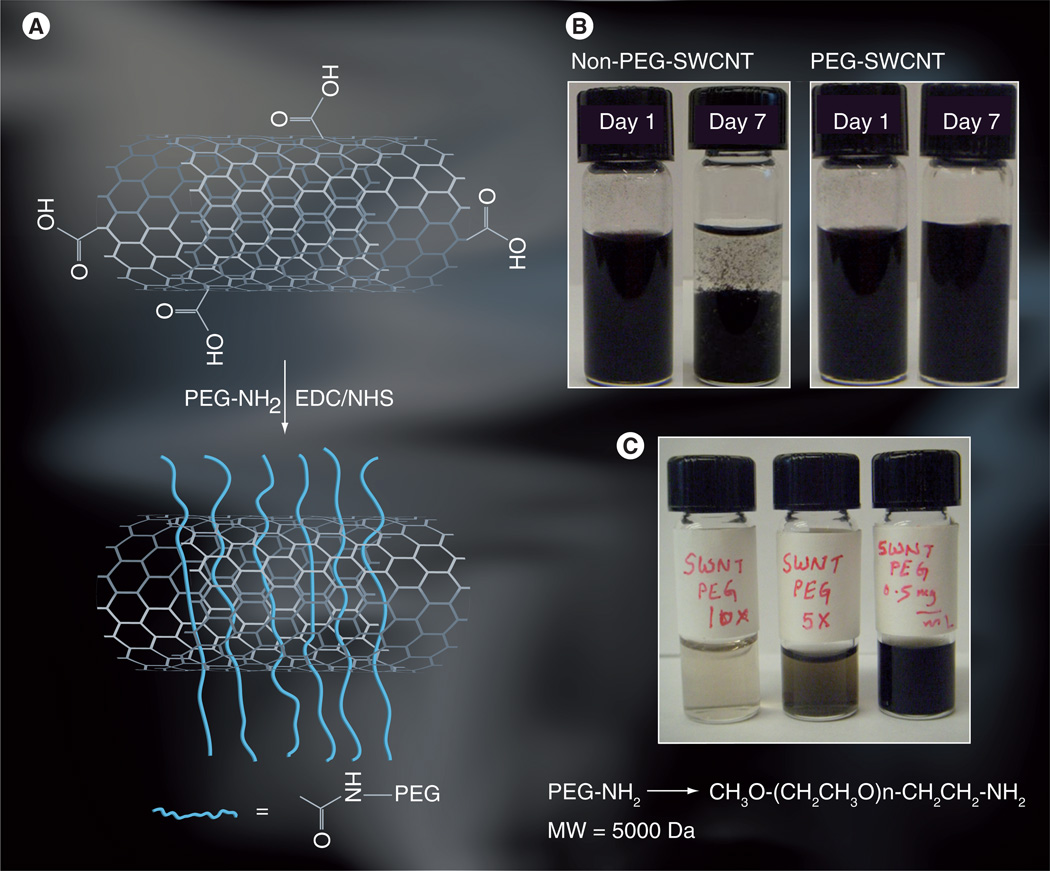
Aims: To study the distribution and clearance of polyethylene glycol (PEG)-ylated single-walled carbon nanotube (SWCNTs) as drug delivery vehicles for the anticancer drug cisplatin in mice.

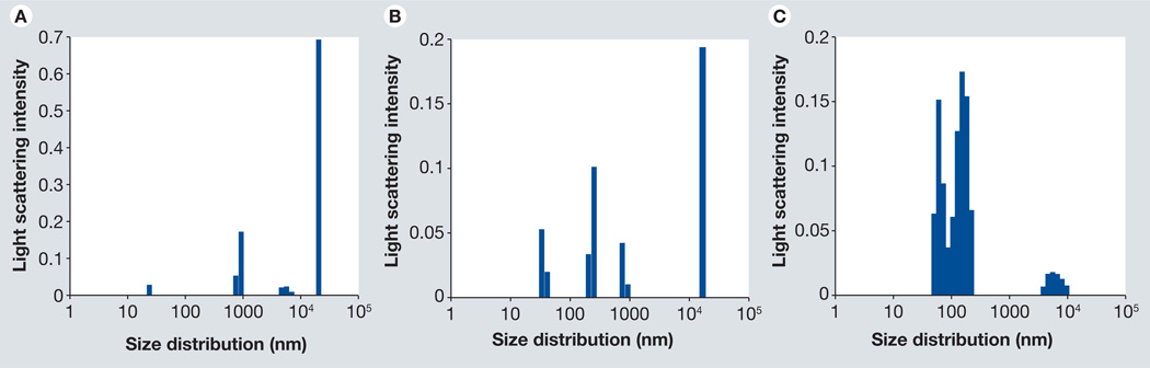
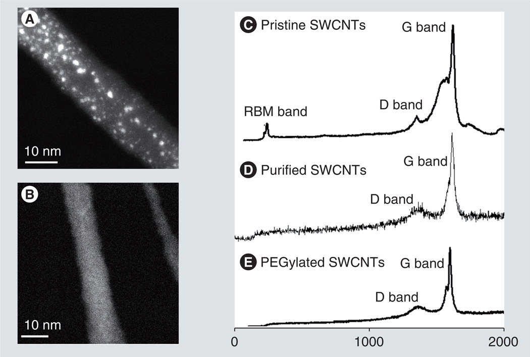
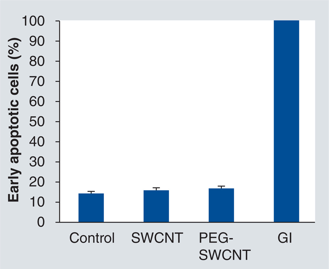
Materials & methods: PEG layers were attached to SWCNTs and dispersed in aqueous media and characterized using dynamic light scattering, scanning transmission electron microscopy and Raman spectroscopy. Cytotoxicity was assessed in vitro using Annexin-V assay, and the distribution and clearance pathways in mice were studied by histological staining and Raman spectroscopy. Efficacy of PEG-SWCNT-cisplatin for tumor growth inhibition was studied in mice.

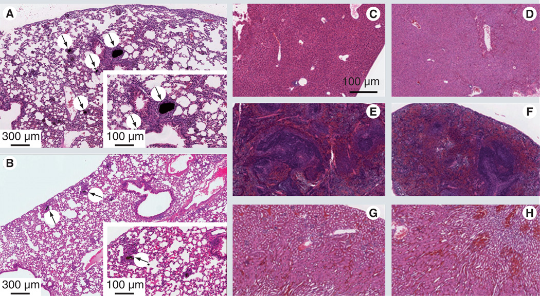
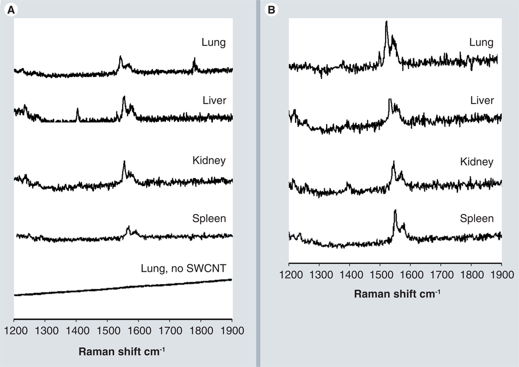
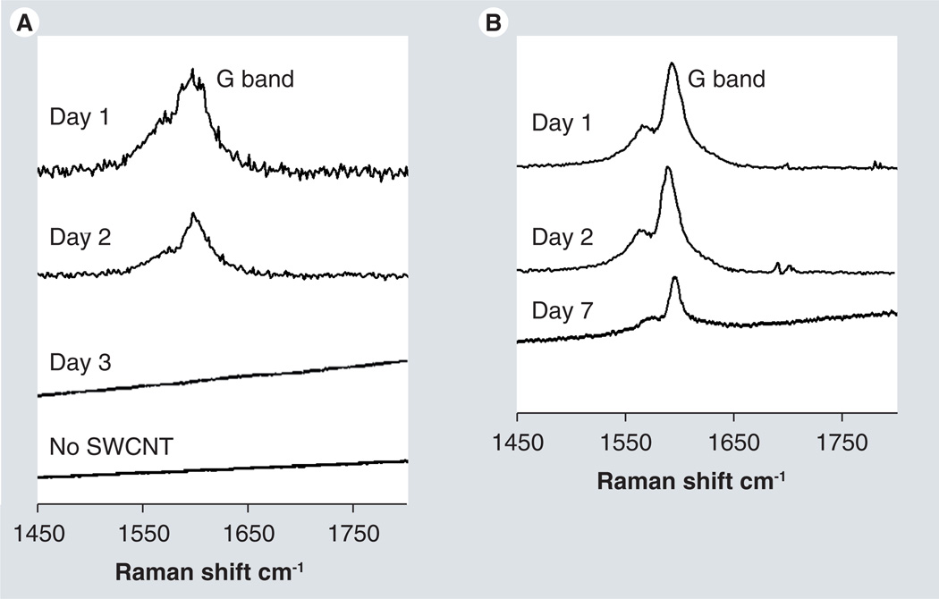
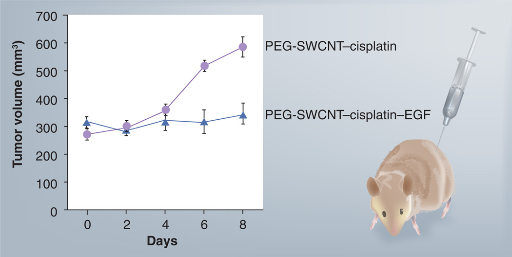
Results & discussion: PEG-SWCNTs were efficiently dispersed in aqueous media compared with controls, and did not induce apoptosis in vitro. Hematoxylin and eosin staining, and Raman bands for SWCNTs in tissues from several vital organs from mice injected intravenously with nanotube bioconjugates revealed that control SWCNTs were lodged in lung tissue as large aggregates compared with the PEG-SWCNTs, which showed little or no accumulation. Characteristic SWCNT Raman bands in feces revealed the presence of bilary or renal excretion routes. Attachment of cisplatin on bioconjugates was visualized with Z-contrast scanning transmission electron microscopy. PEG-SWCNT-cisplatin with the attached targeting ligand EGF successfully inhibited growth of head and neck tumor xenografts in mice.

Conclusions: PEG-SWCNTs, as opposed to control SWCNTs, form more highly dispersed delivery vehicles that, when loaded with both cisplatin and EGF, inhibit growth of squamous cell tumors.


Conflict of interest statement

Financial & competing interests disclosure

The authors have no other relevant affiliations or financial involvement with any organization or entity with a financial interest in or financial conflict with the subject matter or materials discussed in the manuscript apart from those disclosed.

No writing assistance was utilized in the production of this manuscript.

Figures

Figure 1
Figure 1. PEGylated single-walled carbon nanotube attachment and dispersions
(A) Oxidized SWCNT attachment by amide linkage to amino-PEG providing very high dispersion. (B) Photographs of PEG-SWCNT dispersion 1 and 7 days after derived, showing the stability of these nanotube dispersions. Precipitation was seen for non-PEG-SWCNT dispersions. (C) Photographs of PEG-SWCNT dispersions in phosphate buffer saline at various dilutions showing stable dispersions are obtained at higher dilutions. EDC: 1-[3-(dimethylamino) propyl]-3-ethylcarbodiimide hydrochloride; MW: Molecular weight; NHS: N-hydroxysuccinimide; PEG: Polyethylene glycol; SWCNT/SWNT: Single-walled carbon nanotube.
Figure 2
Figure 2. Nominal size distributions of PEGylated single-walled carbon nanotubes dispersed in water from dynamic light scattering
(A) Pristine single-walled carbon nanotube (SWCNT) show a high degree of aggregation in aqueous media along with severe polydispersity. (B) Oxidized SWCNTs show somewhat less aggregation compared with pristine SWCNTs, but still have considerable polydispersity. (C) Polyethylene glycol-SWCNT dispersion shows one major distribution with a smaller secondary one, suggesting less aggregation compared with pristine and oxidized SWCNTs.
Figure 3
Figure 3. Z-contrast scanning transmission electron microscopy images and Raman spectra of PEGylated single-walled carbon nanotubes
(A) Scanning transmission electron microscopy image of a PEG-SWCNT bundle conjugated with anticancer drug cisplatin. Bright dots correspond to clusters of Pt atoms (i.e., cisplatin molecules). (B) Scanning transmission electron microscopy image of a PEG-SWCNT bundle alone shows no bright dots. (C–E) Raman spectra showing signature peaks (relative intensities) of (C) pristine SWCNTs, (D) purified or acid-treated SWCNTs, and (E) SWCNTs wrapped with PEG. PEG: Polyethylene glycol; RBM: Radial breathing mode; SWCNT: Single-walled carbon nanotube.
Figure 4
Figure 4. Apoptosis assays of HN12 head and neck cancer cells treated with single-walled carbon nanotubes and PEGylated single-walled carbon nanotubes
Analysis by the Annexin-V assay and subsequent flow cytometry revealed no significant deviation of the percentage of early-stage apoptotic cells among the experimental groups and negative control. Conversely, approximately 100% of the cells irradiated with γ-radiation were apoptotic, serving as a positive control. Concentrations of SWCNT and PEG-SWCNT were 0.5 mg ml−1. The assay was performed 24 h postapplication. Control: Cells only; GI: γ-irradiation control; PEG: Polyethylene glycol; SWCNT: Single-wall carbon nanotube.
Figure 5
Figure 5. Histological analysis of vital organs of mice treated with single-walled carbon nanotubes and PEGylated single-walled carbon nanotubes
Tissue samples were procured from 6-week-old athymic mice that were injected intravenously with 200 µl of 0.5 mg ml−1 of single-walled carbon nanotube (SWCNT) (A, C, E & G) and polyethylene glycol (PEG)-SWCNT (B, D, F & H) and stained with hematoxylin–eosin. (A) Lung parenchyma showing dark, particulate material, which appears to be free and densely packed, or contained in the cytoplasm of macrophages. The accumulations do not seem to have relations with the airways, but rather appear in the interior of blood vessels that have been clogged by the concretions. Besides the macrophages, other inflammatory mononuclear cells are accumulated surrounding the affected areas. (B) The PEG-SWCNT lungs shows much smaller and fewer concretions similar to those described above. They are also surrounded by mononuclear reactive cells, most likely macrophages. Isolated (segmental) angiocentric chronic inflammatory infiltrates are also present. (C & D) Liver parenchyma with no apparent histological signs of toxicity. (E & F) The kidney has no noticeable histological signs of nanotube toxicity. (G & H) Spleen tissue with no significant histological changes.
Figure 6
Figure 6. Raman analysis of tissue from vital organs
(A) Raman spectra of tissues from vital organs removed from the mice after being treated with polyethylene glycol (PEG)-single-walled carbon nanotubes (SWCNTs). Mice treated with PEG-SWCNTs 24 h post intravenous injection, tissue removed 24 h post intravenous injection for Raman analysis with control lung tissue. The Raman G band for SWCNTs in the 1500–1600 cm−1 region is clearly observed in mice treated with PEG-SWCNTs, while spectral analysis of tissues of mice not treated with SWCNT (bottom trace) showed no indication of the characteristic G band. (B) Raman spectra of tissues of vital mouse organs after being treated with non-PEG-SWCNTs, 24 h post intravenous injection. All spectra are the average of three spots from a given tissue slide.
Figure 7
Figure 7. Raman analysis of powdered fecal samples
(A) Raman spectra of excreted material from mice treated with SWCNTs (days 1–3) or phosphate buffer (no SWCNT). Spectra shown are the average of three spots from the tissue slide. (B) Raman spectra of excreted material from mice treated with polyethylene glycol-SWCNTs treated mice at indicated times (1–7 days). Spectra are the average of three spots from a slide on which the powdered samples were spread. SWCNT: Single-walled carbon nanotube.
Figure 8
Figure 8. Inhibition of pre-established HN12 head and neck squamous carcinoma xenografts by PEG-SWCNTs–cisplatin–EGF
A single injection of the two bioconjugates (PEG-SWCNT–cisplatin with and without EGF 0.5 mg ml−1; approximately 1.33 µM cisplatin concentration) was administered intravenously as shown in the schematic (right) and lesions were measured every 2 days for tumor volume (left). Error bars represent n = 3. PEG: Polyethylene glycol; SWCNT: Single-walled carbon nanotube.

References

    1. Hirsch A. Functionalization of single-walled carbon nanotubes. Angew. Chem. Int. Ed. 2002;41:1853–1859. - PubMed
    1. Bianco A, Kostarelos K, Prato M. Opportunities and challenges of carbon-based nanomaterials for cancer therapy. Expert Opin. Drug Deliv. 2008;5:331–342. •• Describes the biomedical applications of a variety of carbon nanomaterials studied, especially nanotubes. Discusses the potential benefits and risks of carbon nanomaterials towards clinical application.

    1. Dillon AC, Yudasaka M, Dresselhaus MS. Employing Raman spectroscopy to qualitatively evaluate the purity of carbon single-wall nanotube materials. J. Nanosci. Nanotechnol. 2004;4:691–703. - PubMed
    1. Kam S, Dai H. Carbon nanotubes as intracellular protein transporters. generality and biological functionality. J. Am. Chem. Soc. 2005;127:6021–6026. - PubMed
    1. Yang R, Yang X, Zhang Z, et al. Single-walled carbon nanotubes-mediated in vivo and in vitro delivery of siRNA into antigen-presenting cells. Gene Therapy. 2006;13:1714–1723. - PubMed

Publication types