

A local increase in red blood cell aggregation can trigger deep vein thrombosis: evidence based on quantitative cellular ultrasound imaging
- PMID: 21143377
- PMCID: PMC3050084
- DOI: 10.1111/j.1538-7836.2010.04164.x
A local increase in red blood cell aggregation can trigger deep vein thrombosis: evidence based on quantitative cellular ultrasound imaging
Abstract
Background: Recurrent deep vein thrombosis (DVT) risk factors include a first idiopathic DVT, strongly suggesting the existence of undiagnosed and/or unidentified prothrombotic abnormalities.
Objectives: To evaluate the impact of locally increased red blood cell (RBC) aggregation on DVT pathogenesis in a rabbit model.
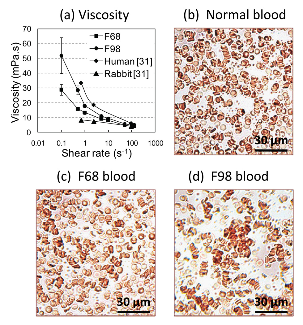
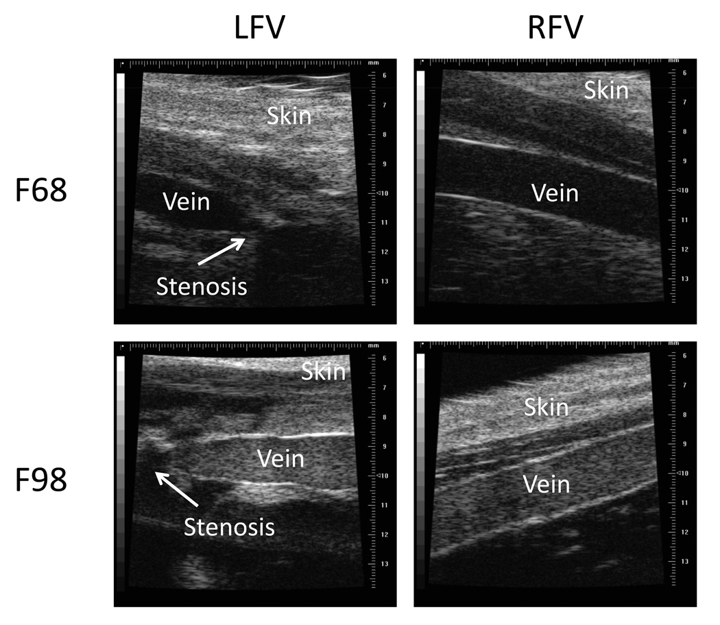
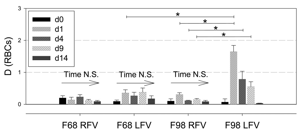
Methods: DVT presence, flow and aggregation were measured in situ with ultrasound. Greatly enhanced aggregation was achieved by covalent linkage of Pluronic F98 to the RBC surface; coating with Pluronic F68, which very mildly enhances aggregation, was used as a coating control. On day 1, endothelial damage and a partial stenosis were surgically created on the left femoral vein whereas the right femoral vein was not manipulated.
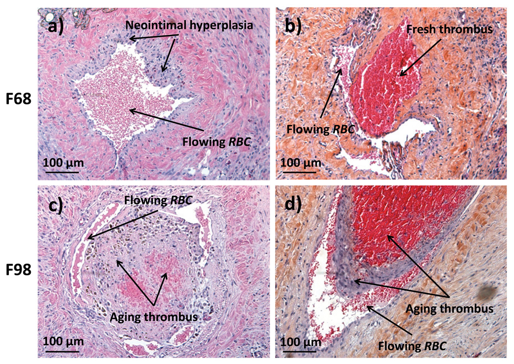
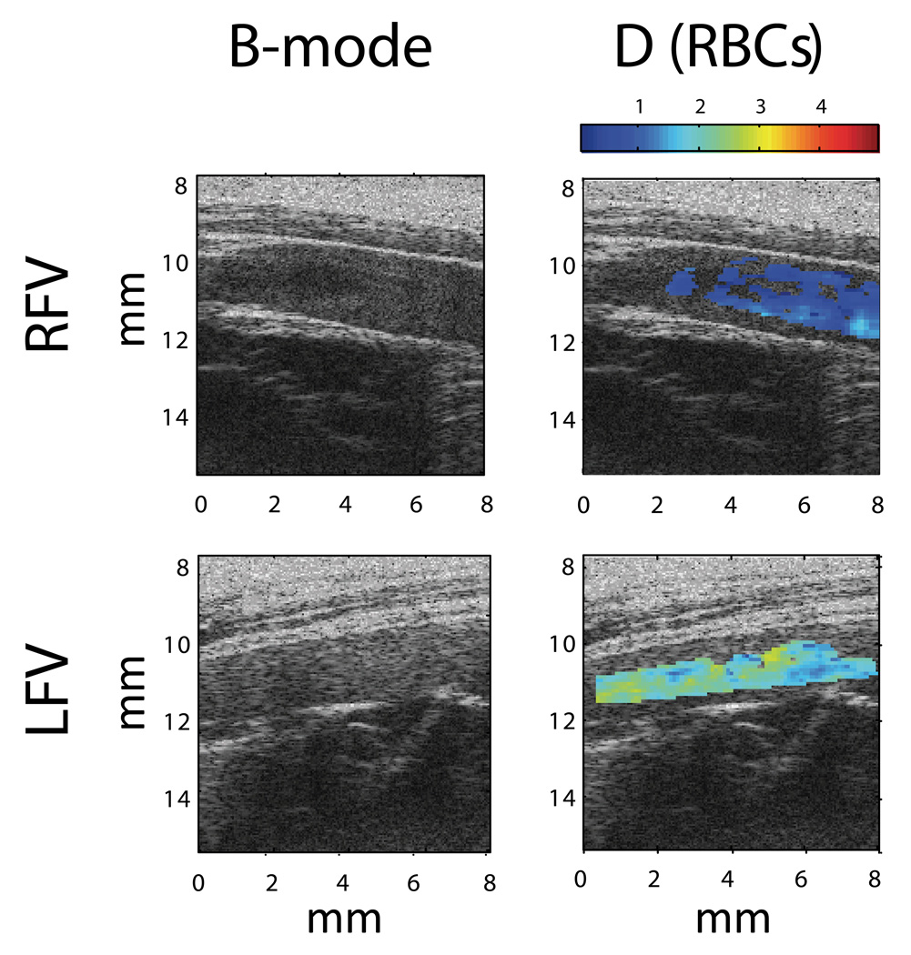
Results: A thrombus was formed within 30 min in six out of seven left femoral veins of animals receiving a 30% volume blood exchange with F98-coated RBC, whereas a thrombus occurred in only one out of seven veins in F68-transfused controls. In vivo imaging using quantitative ultrasound confirmed increased aggregation in the thrombosed veins of the F98 group compared with the F68 group and the contralateral vessel. For each group, five animals were followed for 2 weeks before being killed. In F98-transfused animals, lysis of clots occurred and the presence of chronic thrombi totally occluding the vein in three out of five animals was confirmed by histology. Conversely, in the F68 group, a single disorganized blood clot was observed in one out of five animals.
Conclusions: A marked increase in RBC aggregation promotes thrombosis in rabbit femoral veins, confirming a pathophysiological role of locally altered hemorheology in the onset of DVT.
© 2011 International Society on Thrombosis and Haemostasis.
Figures
References
-
- Agnelli G, Becattini C. Treatment of DVT: how long is enough and how do you predict recurrence. J Thromb Thrombolysis. 2008;25:37–44. - PubMed
-
- Alt E, Banyai S, Banyai M, Koppensteiner R. Blood rheology in deep venous thrombosis--relation to persistent and transient risk factors. Thromb Res. 2002;107:101–107. - PubMed
-
- Vaya A, Mira Y, Martinez M, Villa P, Ferrando F, Estelles A, Corella D, Aznar J. Biological risk factors for deep vein trombosis. Clin Hemorheol Microcirc. 2002;26:41–53. - PubMed
-
- Christiansen SC, Cannegieter SC, Koster T, Vandenbroucke JP, Rosendaal FR. Thrombophilia, clinical factors, and recurrent venous thrombotic events. JAMA. 2005;293:2352–2361. - PubMed
-
- Schulman S, Ogren M. New concepts in optimal management of anticoagulant therapy for extended treatment of venous thromboembolism. Thromb Haemost. 2006;96:258–266. - PubMed
Publication types
MeSH terms
Substances
Grants and funding
LinkOut - more resources
Full Text Sources
Medical
