

Erdheim-Chester disease presenting with cutaneous involvement: a case report and literature review
- PMID: 21143617
- PMCID: PMC3619727
- DOI: 10.1111/j.1600-0560.2010.01650.x
Erdheim-Chester disease presenting with cutaneous involvement: a case report and literature review
Abstract
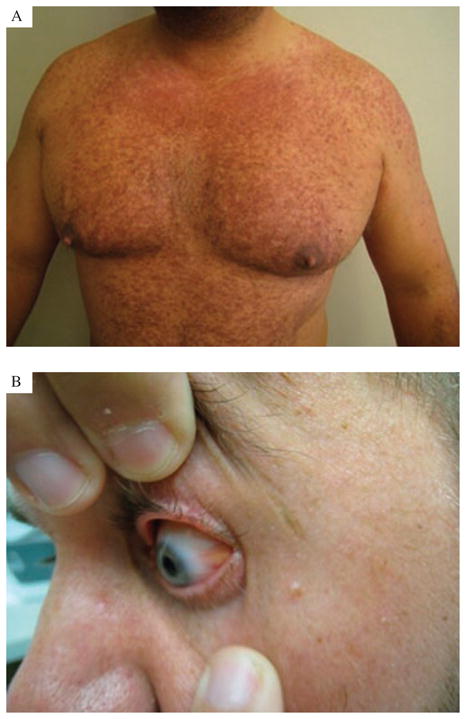
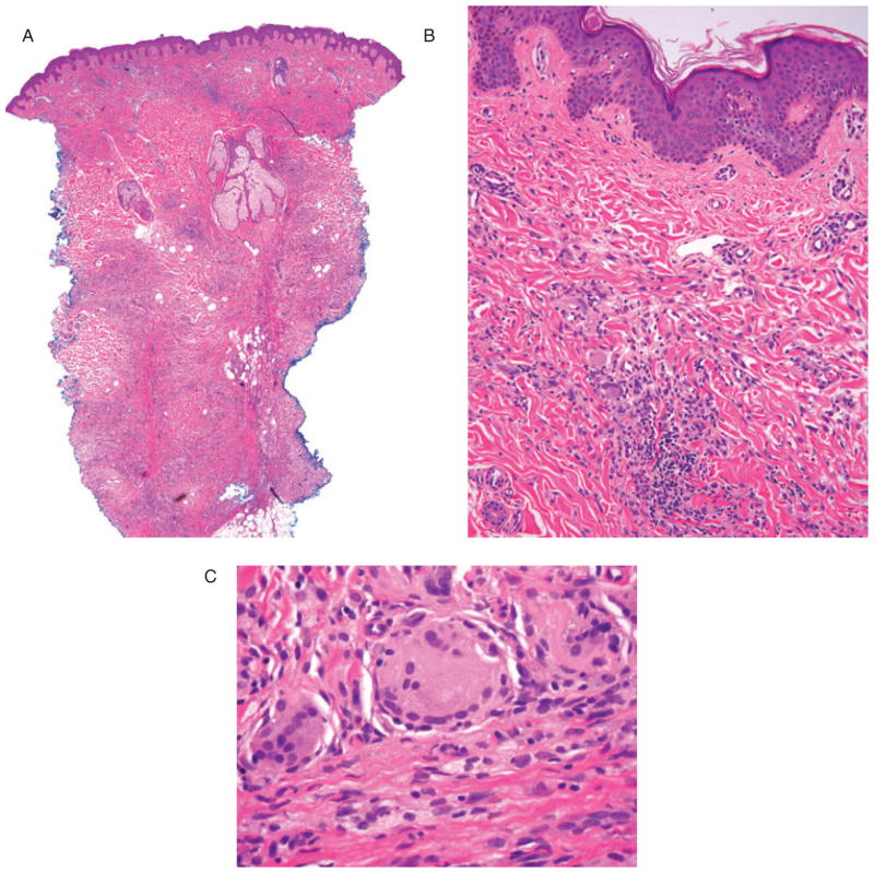
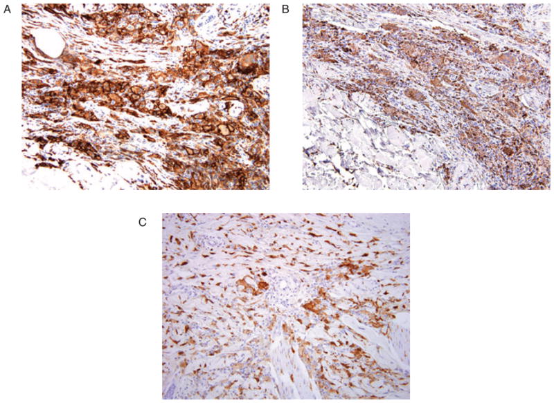
Erdheim-Chester disease (ECD) is a rare, systemic, non-familial histiocytic disorder, first described by Jakob Erdheim and William Chester in 1930. Most patients have multiple sites of involvement at presentation. The most common site of involvement is the long bones of the axial skeleton, which is seen almost universally, followed by the nervous system, heart, lungs, orbit and retroperitoneum, which are seen in up to 50% of cases. Cutaneous involvement is rarely a presenting symptom of ECD, with two reported cases in the English literature. The diagnosis of ECD is rarely made by skin biopsy because of the relative rarity of cutaneous involvement as a presenting feature, and also perhaps because of the difficulty in distinguishing the histopathological appearance from potential mimics. The importance of distinguishing ECD from other cutaneous disorders with similar pathology lies in the implications for both treatment and prognosis. ECD is an aggressive, often fatal disorder, with death from disease occurring in greater than 50% of patients.
Copyright © 2010 John Wiley & Sons A/S.
Figures
References
-
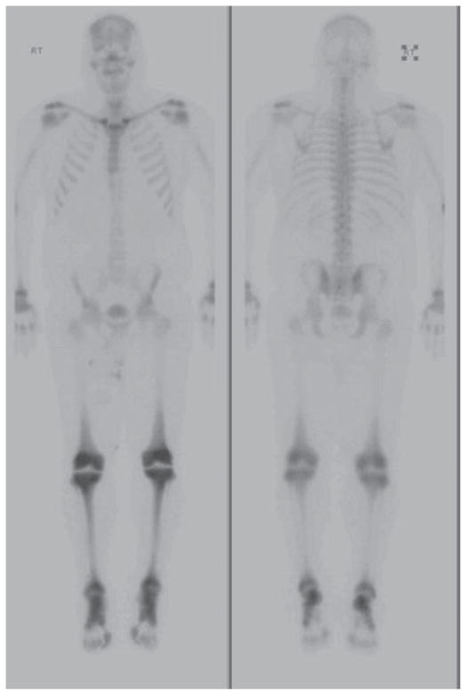
- Spyridonidis TJ, Giannakenas C. Erdheim-Chester Disease: a rare syndrome with a characteristic bone scinitigraphy pattern. Ann Nucl Med. 2008;22:323. - PubMed
-
- Garg T, Chander R, Gupta T, Mendiratta V, Jain M. Erdheim-Chester disease with cutaneous features in an Indian patient. Skinmed. 2008;7:103. - PubMed
-
- Vencio EF, Jenkins RB. Clonal cytogenetic abnormalities in Erdheim-Chester disease. Am J Surg Pathol. 2007;31:319. - PubMed
-
- Al-Quran S, Reith J, Bradley J, Rimsza L. Erdheim-Chester disease: case report, PCR-based analysis of clonality, and review of literature. Mod Pathol. 2002;15:666. - PubMed
-
- Veyssier-Belot C, Cacoub P. Erdheim-Chester disease. Clinical and radiologic characteristics of 59 cases. Medicine (Balitimore) 1996;75:157. - PubMed
Publication types
MeSH terms
Grants and funding
LinkOut - more resources
Full Text Sources
Medical
