. 2010 Dec 8;11(1):174.
doi: 10.1186/1465-9921-11-174.

Population specificity of the DNAI1 gene mutation spectrum in primary ciliary dyskinesia (PCD)

Affiliations

Population specificity of the DNAI1 gene mutation spectrum in primary ciliary dyskinesia (PCD)

Ewa Ziętkiewicz et al. Respir Res. .

Abstract

Background: Mutations in the DNAI1 gene, encoding a component of outer dynein arms of the ciliary apparatus, are the second most important genetic cause of primary ciliary dyskinesia (PCD), the genetically heterogeneous recessive disorder with the prevalence of ~1/20,000. The estimates of the DNAI1 involvement in PCD pathogenesis differ among the reported studies, ranging from 4% to 10%.

Methods: The coding sequence of DNAI1 was screened (SSCP analysis and direct sequencing) in a group of PCD patients (157 families, 185 affected individuals), the first ever studied large cohort of PCD patients of Slavic origin (mostly Polish); multiplex ligation-dependent probe amplification (MLPA) analysis was performed in a subset of ~80 families.

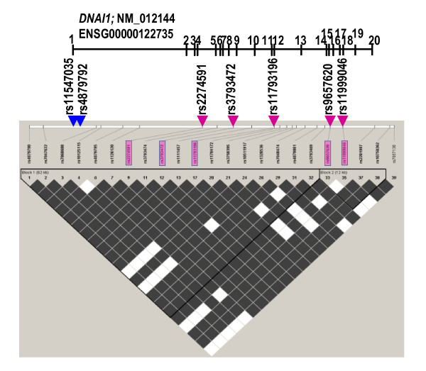
Results: Three previously reported mutations (IVS1+2-3insT, L513P and A538T) and two novel missense substitutions (C388Y and G515S) were identified in 12 families (i.e. ~8% of non-related Polish PCD patients). The structure of background SNP haplotypes indicated common origin of each of the two most frequent mutations, IVS1+2-3insT and A538T. MLPA analysis did not reveal any significant differences between patients and control samples. The Polish cohort was compared with all the previously studied PCD groups (a total of 487 families): IVS1+2-3insT remained the most prevalent pathogenetic change in DNAI1 (54% of the mutations identified worldwide), and the increased global prevalence of A538T (14%) was due to the contribution of the Polish cohort.

Conclusions: The worldwide involvement of DNAI1 mutations in PCD pathogenesis in families not preselected for ODA defects ranges from 7 to 10%; this global estimate as well as the mutation profile differs in specific populations. Analysis of the background SNP haplotypes suggests that the increased frequency of chromosomes carrying A538T mutations in Polish patients may reflects local (Polish or Slavic) founder effect. Results of the MLPA analysis indicate that no large exonic deletions are involved in PCD pathogenesis.


Figures

Figure 1
Figure 1
Characteristics of three new variants detected in PCD patients. a. Results of the SSCP analysis revealing different migration patterns, and pedigrees of the families where new mutations/SNPs were identified. New mutations (in patients 537, in his father 538, and in patients 507 and 336) are underlined. "?" denotes unknown mutation; the carrier status is indicated by a dot in the pedigree symbols. b. Chromatograms of the new sequence variants.
Figure 2
Figure 2
Evolutionary conservation of the sites of two missense mutations. The positions of two missense mutations (arrows) with respect to the position of WD-blocks 2 and 4 (boxed) in exons 13 and 16-17; species comparison indicates high evolutionary conservation.
Figure 3
Figure 3
Substitution in the DNAI1 3'UTR region. The position of the substitution is highlighted; putative regulatory motifs in the DNAI1 3'UTR region are underlined.
Figure 4
Figure 4
Distribution of the 7-position SNP haplotype variants among the studied chromosomes. Positions of the pathogenic mutations are indicated in on the SNP haplotype background. H1 through H8r2 are arbitrary names of the variants of the 7-position SNP haplotype. SNP1, 2, 3, 4, 5, 6 and 7 denote, respectively, rs11547035, rs4879792, rs2274591, rs3793472, rs11793196, rs9657620, rs11999046. Letters "0" and "1" in the left section of this Figure indicate, respectively, the ancestral and derived allele of the SNPs (the ancestral alleles were determined from the human-chimpanzee comparison, with the sequence identity indicating the ancestral state). Minimal regions of recombination (letter "r" in the haplotype name) in the rare haplotype variants, proposed assuming most parsimonious recombination among the frequent variants, and taking into account the extent of LD in the gene region (Figure 5), are highlighted. Right section of the Figure indicates the number of chromosomes with the respective haplotype variants. *G > A at rs11793196 and c.1612 are transitions at CpG dinucleotides. Mutation A538T on the H1 background was found only in KS families; A538T on the H8r2 was found in a CDO family. Recombination detected within the PCD family. § "0-1" at the last position of the H6 haplotype denotes ancestral allele (G) at rs11999046, linked with the derived allele (A) 93 nt downstream from rs11999046. **Unknown mutation(s) in two PCD families.
Figure 5
Figure 5
Linkage disequilibrium (LD) across the DNAI1 gene. Based on the HapMapCEU SNPs triangle plot generated for HapMap CEU data (release 21) for ENSG00000122735 (chrom 9:34457-34521 kb) with the use of Haploview software. Positions of SNPs genotyped in PCD families are indicated by arrowheads (rs11547035 and rs4879792, blue arrowheads, are not among SNPs from the HapMapCEU panel). The strength of the LD between SNPs (solid spine of LD) is indicated by colors: white (low) and dark (high); LD blocks are indicated by black triangles. Haplotypes flanked by markers rs11547036 and rs11793196 are within a single block of LD.

References

    1. Afzelius BA, Mossberg B. The Metabolic and Molecular Bases of Inherited Disease. New York: McGraw-Hill, Inc; 1995. Immotile-cilia syndrome (primary ciliary dyskinesia), including Kartagener syndrome; pp. 3943–54.
    1. Morillas HN, Zariwala M, Knowles MR. Genetic causes of bronchiectasis: primary ciliary dyskinesia. Respiration. 2007;74:252–63. doi: 10.1159/000101783. - DOI - PubMed
    1. Narayan D, Krishnan SN, Upender M, Ravikumar TS, Mahoney MJ, Dolan TF Jr, Teebi AS, Haddad GG. Unusual inheritance of primary ciliary dyskinesia (Kartagener's syndrome) J Med Genet. 1994;31:493–6. doi: 10.1136/jmg.31.6.493. - DOI - PMC - PubMed
    1. Krawczyński MR, Witt M. PCD and RP: X-linked inheritance of both disorders? Pediatr Pulmonol. 2004;38:88–9. - PubMed
    1. Krawczyński MR, Dmeńska H, Witt M. Apparent X-linked primary ciliary dyskinesia associated with retinitis pigmentosa and a hearing loss. J Appl Genet. 2004;45:107–10. - PubMed

Publication types