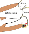
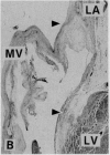
Mitral annular disjunction in myxomatous mitral valve disease: a relevant abnormality recognizable by transthoracic echocardiography
- PMID: 21143934
- PMCID: PMC3014886
- DOI: 10.1186/1476-7120-8-53
Mitral annular disjunction in myxomatous mitral valve disease: a relevant abnormality recognizable by transthoracic echocardiography
Abstract
Background: Mitral annular disjunction (MAD) consists of an altered spatial relation between the left atrial wall, the attachment of the mitral leaflets, and the top of the left ventricular (LV) free wall, manifested as a wide separation between the atrial wall-mitral valve junction and the top of the LV free wall. Originally described in association with myxomatous mitral valve disease, this abnormality was recently revisited by a surgical group that pointed its relevance for mitral valve reparability. The aims of this study were to investigate the echocardiographic prevalence of mitral annular disjunction in patients with myxomatous mitral valve disease, and to characterize the clinical profile and echocardiographic features of these patients.
Methods: We evaluated 38 patients with myxomatous mitral valve disease (mean age 57 ± 15 years; 18 females) and used standard transthoracic echocardiography for measuring the MAD. Mitral annular function, assessed by end-diastolic and end-systolic annular diameters, was compared between patients with and without MAD. We compared the incidence of arrhythmias in a subset of 21 patients studied with 24-hour Holter monitoring.
Results: MAD was present in 21 (55%) patients (mean length: 7.4 ± 8.7 mm), and was more common in women (61% vs 38% in men; p = 0.047). MAD patients more frequently presented chest pain (43% vs 12% in the absence of MAD; p = 0.07). Mitral annular function was significantly impaired in patients with MAD in whom the mitral annular diameter was paradoxically larger in systole than in diastole: the diastolic-to-systolic mitral annular diameter difference was -4,6 ± 4,7 mm in these patients vs 3,4 ± 1,1 mm in those without MAD (p < 0.001). The severity of MAD significantly correlated with the occurrence of non-sustained ventricular tachycardia (NSVT) on Holter monitoring: MAD›8.5 mm was a strong predictor for (NSVT), (area under ROC curve = 0.74 (95% CI, 0.5-0.9); sensitivity 67%, specificity 83%). There were no differences between groups regarding functional class, severity of mitral regurgitation, LV volumes, and LV systolic function.
Conclusions: MAD is a common finding in myxomatous mitral valve disease patients, easily recognizable by transthoracic echocardiography. It is more prevalent in women and often associated with chest pain. MAD significantly disturbs mitral annular function and when severe predicts the occurrence of NSVT.
Figures


References
-
- Eriksson MJ, Bitkover CY, Omran AS, David T, David TE, Ivanov J, Ali MJ, Woo A, Siu SC, Rakowski H. Mitral annular disjunction in advanced myxomatous mitral valve disease: Echocardiographic detection and surgical correction. J Am Soc Echocardiogr. 2005;18:1014–1022. doi: 10.1016/j.echo.2005.06.013. - DOI - PubMed
-
- Playford D, Weyman AE. Mitral valve prolapse: time for fresh look. Rev Cardiovasc Med. 2001;2:73–81. - PubMed
-
- Lang RM, Bierig M, Devereux RB, Flachskampf FA, Foster E, Pellikka PA, Picard MH, Roman MJ, Seward J, Shanewise JS, Solomon SD, Spencer KT, Sutton MS, Stewart WJ. Recommendations for chamber quantification: A report from the ASE's guidelines and standards committee and the chamber quantification writing group, developed in conjunction with the ESE, a branch of the ESC. Eur J Echocardiogr. 2006;7:79–108. doi: 10.1016/j.euje.2005.12.014. - DOI - PubMed
-
- Lancellotti P, Moura L, Pierard LA, Agricola E, Popescu BA, Tribouilloy C, Hagendorff A, Monin JL, Badano L, Zamorano JL. European Association of Echocardiography recommendations for the assessment of valvular regurgitation. Part 2: mitral and tricuspid regurgitation (native valve disease) Eur J Echocardiogr. 2010;11:307–332. doi: 10.1093/ejechocard/jeq031. - DOI - PubMed
MeSH terms
LinkOut - more resources
Full Text Sources
Molecular Biology Databases

