. 2010 Dec 13;9 Suppl 3(Suppl 3):S6.
doi: 10.1186/1475-2875-9-S3-S6.

Apoptosis stalks Plasmodium falciparum maintained in continuous culture condition

Affiliations

Apoptosis stalks Plasmodium falciparum maintained in continuous culture condition

Beth K Mutai et al. Malar J. .

Abstract

Background: Growth kinetic of Plasmodium falciparum in culture or in the host fall short of expected growth rate considering that there are 4 x 10(6)/µL red blood cell (RBCs) available for invasion and about 16 merozoites growing in each infected RBC. This study determined whether apoptotic machinery is operable to keep the parasite population under check.

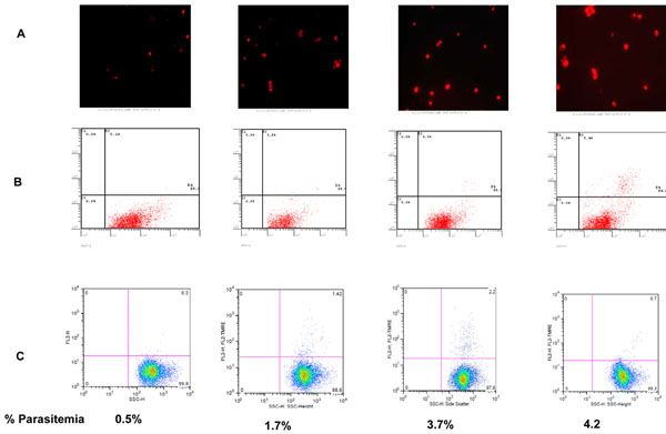
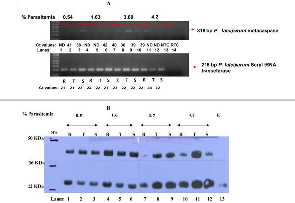
Methods: A synchronized culture of P. falciparum (Dd2 strain) was initiated at 0.5% ring stage parasitaemia and kept under conditions not limiting for RBCs and nutrient by adjusting hematocrit to 5% at each schizogony and changing growth media daily. Parasite growth pattern and morphology was evaluated by blood smear microscopy and flow-cytometry using SYBR green. The apoptotic processes were evaluated for evidence of: DNA fragmentation by TUNEL, collapse of mitochondria membrane potential (ΔΨm) by TMRE, expression of metacaspase gene by RT-qPCR and by probing parasite proteins with anti-caspase antibodies.

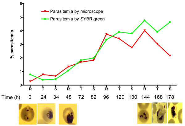
Results: From the seeding parasitaemia of 0.5%, the parasites doubled every 48 hours to a parasitaemia of 4%. Thereafter, the growth stagnated and the culture consistently crashed at about 6% parasitaemia. ΔΨm potential collapsed as the parasite density increased and DNA fragmentation increased steadily from 0.2% to ~6%. The expression of metacaspase gene and protein was observed in all stages, but their abundance was variable among the stages.

Conclusion: These findings suggest existence of P. falciparum quorum sensing that keep the parasite population under check.


Figures

Figure 1
Figure 1
In vitro growth pattern of P. falciparum Dd2 isolate as measured by microscopy (red curve and photomicrograph) and SYBR Green (green curve). By both methods, parasitaemia initiated at 0.5% doubled at every schizogony and did not increase beyond 5%. The photomicrograph show healthy parasite at seeding parasitaemia: note the classical signet ring, healthy looking trophozoites with irregular borders and mature schizonts with numerous nuclei. At maximal parasitaemia, the ring stages were smaller with intensely stained nuclei while the trophozoites and schizonts have abnormal nuclei.
Figure 2
Figure 2
Panel A and B: Level of fragmented DNA using terminal deoxynucleotidyl-transferase-mediated dUTP nick-end labeling (TUNEL). Evidence of DNA fragmentation as measured by TUNEL is discernible albeit at very low level by fluorescent microscopy (Panel A) and flow cytometry (Panel B) at seeding parasitaemia, but increases dramatically by the time the parasite culture crashes. Panel C: Dot blots showing percentage accumulation of TMRE in the mitochondria of trophozoites at different parasitaemia. As shown, the proportion of cells with positive TMRE staining was maximal at 3.7% parasitaemia and declined thereafter.
Figure 3
Figure 3
Panal A: Agarose gel picture showing expression level of metacaspase and Seryl transferase genes in rings (R), trophozoites (T) and schizonts (S) at different parasite densities (double arrow bar). NTC= No template control (Lane 13 ). RTC= Reverse transcription control (lane 14). Expression of metacaspase gene was stage dependent. At the ring stage metacaspase was absent until the parasite density reached 4%. For the trophozoites, the metacaspase gene was detectable at 0.54% and 3.68% parasitaemias while in the schizonts, the gene was present at all parasitaemia levels. The P. falciparum house keeping gene, seryl tRNA, was expressed at relatively equal level in all the RNA preparations. Panel B: Western blot analysis of P. falciparum protein extract probed with anti- caspase-7 polyclonal antibody. Protein fragments of 45 and 28 kDa were observed in all the developmental stages (lanes 1-12). Metacaspase protein was up-regulated starting from parasite density of 1.6% in all the parasite stages and remained up-regulated in subsequent parasitaemia levels except in the ring (lanes 7 and 10). Note a low levels of protein of 28 kDa in uninfected erythrocytes (lane 13) and has been reported before in other negative control samples. [16]

References

    1. Deponte M, Becker K. Plasmodium falciparum--do killers commit suicide? Trends Parasitol. 2004;20(4):165–169. doi: 10.1016/j.pt.2004.01.012. - DOI - PubMed
    1. Ginsburg H, Hoshen MB. Is the development of falciparum malaria in the human host limited by the availability of uninfected erythrocytes? Malar J. 2002;1:18. doi: 10.1186/1475-2875-1-18. - DOI - PMC - PubMed
    1. Domen J. The role of apoptosis in regulating hematopoietic stem cell numbers. Apoptosis. 2001;6(4):239–252. doi: 10.1023/A:1011347623402. - DOI - PubMed
    1. Samali A, Zhivotovsky B, Jones D, Nagata S, Orrenius S. Apoptosis: cell death defined by caspase activation. Cell Death Differ. 1999;6(6):495–496. doi: 10.1038/sj.cdd.4400520. - DOI - PubMed
    1. Alnemri ES, Livingston DJ, Nicholson DW, Salvesen G, Thornberry NA, Wong WW, Yuan J. Human ICE/CED-3 protease nomenclature. Cell. 1996;87(2):171. doi: 10.1016/S0092-8674(00)81334-3. - DOI - PubMed

LinkOut - more resources