. 2011 Feb 1;350(1):127-38.
doi: 10.1016/j.ydbio.2010.11.028. Epub 2010 Dec 7.

Interactions between oligodendrocyte precursors control the onset of CNS myelination

Affiliations

Interactions between oligodendrocyte precursors control the onset of CNS myelination

Yan Yang et al. Dev Biol. .

Abstract

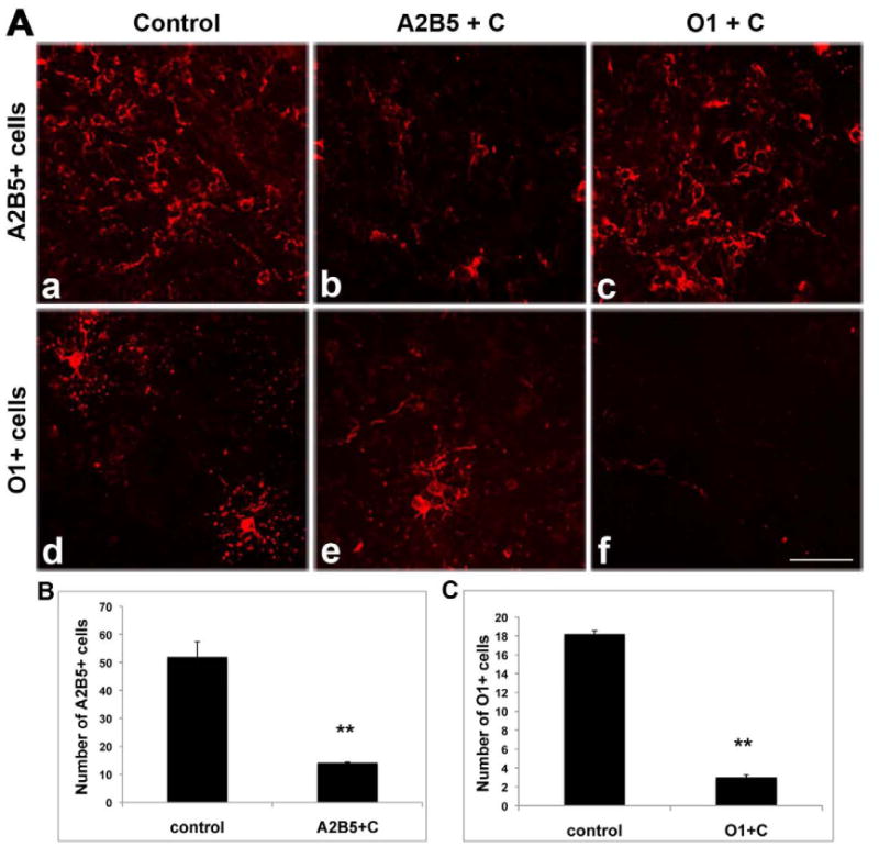
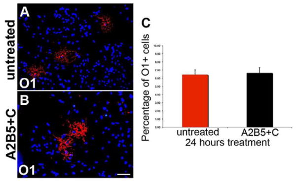
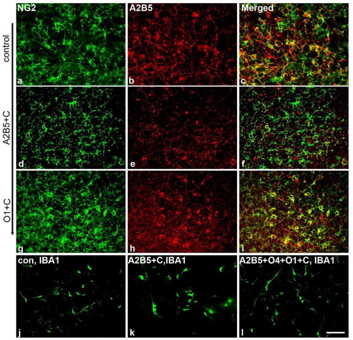
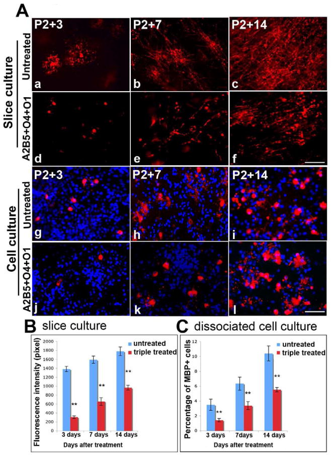
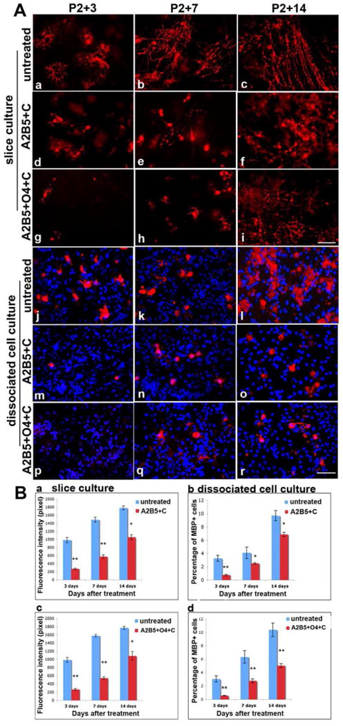
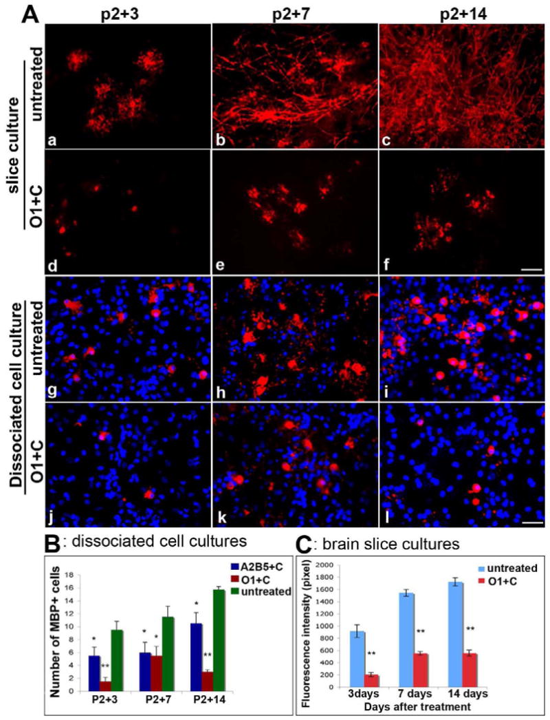
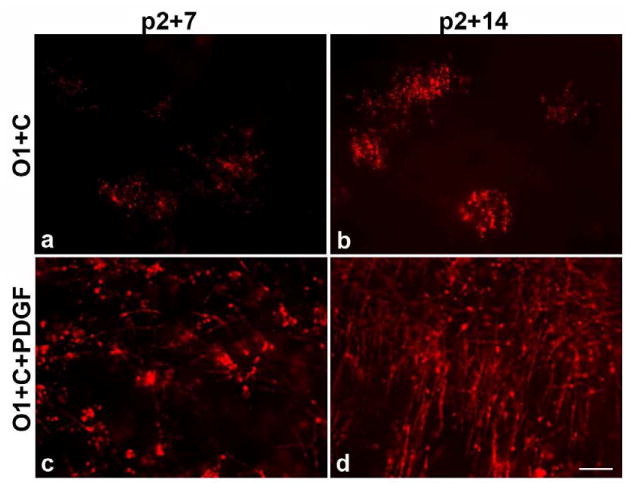
The formation of CNS myelin is dependent on the differentiation of oligodendrocyte precursor cells (OPCs) and oligodendrocyte maturation. How the initiation of myelination is regulated is unclear, but it is likely to depend on the development of competence by oligodendrocytes and receptivity by target axons. Here we identify an additional level of control of oligodendrocyte maturation mediated by interactions between the different cellular components of the oligodendrocyte lineage. During development oligodendrocyte precursors mature through a series of stages defined by labeling with monoclonal antibodies A2B5 and O4. Newly differentiated oligodendrocytes begin to express galactocerebroside recognized by O1 antibodies and subsequently mature to myelin basic protein (MBP)-positive cells prior to formation of compact myelin. Using an in vitro brain slice culture system that supports robust myelination, the consequences of ablating cells at different stages of the oligodendrocyte lineage on myelination have been assayed. Elimination of all OPC lineage cells through A2B5+, O4+, and O1+ complement-mediated cell lysis resulted in a delay in development of MBP cells and myelination. Selective elimination of early OPCs (A2B5+) also unexpectedly resulted in delayed MBP expression compared to controls suggesting that early OPCs contribute to the timing of myelination onset. By contrast, elimination of differentiated (O1+) immature oligodendrocytes permanently inhibited the appearance of MBP+ cells suggesting that oligodendrocytes are critical to facilitate the maturation of OPCs. These data illuminate that the presence of intra-lineage feed-forward and feedback cues are important for timely myelination by oligodendrocytes.


Figures

Figure 1
Figure 1
Development of myelination in rat brain slice cultures. P2 brain coronal slices taken from the region 1.1-02 relative to Bregma were grown in vitro for 3, 5, 7 or 14 days. Immature MBP+ cells with few processes were seen in the developing motor cortex and underlying white matter at P2+3 DIV (a). In the cultures at P2+5 DIV (b) and P2+7 DIV (c), more MBP+ cells with extended processes had developed by P2+14 DIV (d), MBP is expressed both in oligodendrocyte cell bodies and in processes surrounding axons. Black-Gold® staining (e) confirms the presence of myelination in P2+14 DIV slice cultures. The close association of axons and oligodendrocyte process is demonstrated by the high level of coincidence of MBP (red) co-localized with neuronal axons, which is stained by neurofilament (green) in (f). Confocal imaging (g) confirms that axons are wrapped by MBP+ process. Electron microscopy (EM) shows an axon in longitudinal section surrounded by a compacted myelin sheath (arrows) at P2+14 DIV (h). cx = cortex, cc = corpus callosum. Scale bar = 25μm.
Figure 2
Figure 2
Reduction of A2B5+ or O1+ cells in slice cultures after complement-mediated lysis to selectively ablate A2B5+ or O1+ cells. A dramatic reduction in the number of A2B5+ cells (A,b) was seen following treatment with A2B5 plus complement (A2B5+C), compared to the untreated control (A,a) whereas the density of O1+ cells remains at a similar level to control (A,e). Likewise, treatment with O1 antibody plus complement resulted in substantial loss of O1+ cells (A,f) while sparing A2B5+ cells (A,c). Quantitative data are shown in B and C. **p<0.01, treated groups versus controls. Scale bar =25μm
Figure 3
Figure 3
The number of O1+ immature oligodendrocytes remains unchanged before and after complement mediated A2B5+ cell ablation in dissociated cell cultures. A, O1-positive cells were found in untreated primary brain mixed culture after 24 hours. In cultures exposed to A2B5 plus complement mediated lysis (A2B5+C), O1-positive cells are still present (B) and quantification (C) shows that the number of O1-positive cells is not significantly difference in treated and untreated cell cultures (p>0.05). Scale bar = 25μm.
Figure 4
Figure 4
Ablation of A2B5+ cells reduces the density of NG2+ cells while ablation of O1+ cells does not dramatically affect NG2+ cell numbers. Complement mediated cell lysis in slice cultures does not stimulate a widespread microglial response. Control cultures show significant overlap in expression of A2B5 (red) and NG2 (green) on OPCs (a-c). A discrete population of NG2+ cells is also present and is retained following ablation of A2B5+ cells (d-f). Parallel studies with O1 targeted complement lysis do not substantially alter the composition of the pool of OPCs (g-i). To determine whether complement mediated cell lysis stimulated a microglial response, slice cultures were labeled with IBA1 antibodies in controls and after A2B5 or triple antibody mediated cell lysis (j-l). No increase in IBA1 staining obviously accompanied cell lysis. Scale bar = 25μm.
Figure 5
Figure 5
Myelin formation is delayed in the absence of all stages of the committed OPC lineage. Cultured slices were treated with A2B5, O4 and O1 antibodies simultaneously, followed by complement-mediated lysis. While some occasional MBP+ process were detectable, few MBP+ cells (red) were seen P2+3 DIV (A,d) and appeared unhealthy compared to untreated P2+3DIV slices (A,a). In the treated groups, MBP+ cells with few myelinated fibrils appear by P2+7 DIV (A,e) and increase by P2+14 DIV(A,f), but myelination was significantly reduced compared to the non-treated slices at the same ages (A,b-c). Similar results were found in primary dissociated brain mixed cell cultures (A,g-l). Quantitation of MBP fluorescence intensity (pixel) shows significantly decreased MBP intensity in triple-treated slice cultures (P<0.01) compared to untreated slice cultures (B). Quantitation of the percent of MBP+ cells in treated dissociated cell cultures demonstrated a significant decrease (p<0.01) compared to untreated cultures (C). Scale bar =25μm.
Figure 6
Figure 6
Myelination is delayed after depleting early stage OPC progenitors. After depleting early progenitors (A2B5+ cells or A2B5+ and O4+ cells), only a few immature MBP+ cells are seen at P2+ 3 DIV (A, d, g) and at P2+7 DIV(A, e, h). In untreated cultures, MBP+ cells were morphologically healthy and more mature at P2+3 DIV (A, a), and process extension started by P2+7 DIV (A, b). By P2+14 DIV, MBP+ processes appeared in the treated culture (A, f, i), but were delayed and disorganized compared to the untreated slice cultures that contain robust, parallel myelinated fibrils at P2+14 DIV (A, c). Similar results were found in primary brain dissociated mixed cell cultures after ablation of A2B5+ cell alone (A, m-o) or A2B5+ and O4+ cells together (A, p-r). Untreated cell cultures are shown in A, j-l. Quantitative analysis of MBP fluorescence intensity (pixel) and the percentage of MBP positive cells in cultures are shown in B, a-d. After depletion of A2B5+ cells alone or A2B5+ plus O4+ progenitors in slice cultures, the intensity of MBP fluorescence is significantly decreased (**p<0.01) compared to untreated slice cultures (B, a, c). Even at P2+14 DIV, when MBP intensity continues to increase, it is still significantly lower than in untreated slice cultures (*p<0.05). The percentage of MBP-positive cells is significant decreased (**p<0.01) in brain dissociated mix cell cultures (B, b, d). Scale bar = 25μm.
Figure 7
Figure 7
Lack of myelin formation and oligodendrocyte maturation in the absence of O1+ immature oligodendrocytes. In slice cultures (a-f) and dissociated cell cultures (g-i) the maturation of oligodendrocytes is dependent on the presence of O1+ cells. Few MBP+ cell bodies were found at P2+3 DIV (A, d) and P2+7 DIV (A, e) following elimination of O1+ cells. MBP+ processes failed to develop in treated cultures even at P2+14 DIV (A,f). Untreated slice cultures are shown in A, a-c. In mixed cell cultures (g-i) treated with O1 plus complement (O1+C), MBP+ cells were reduced and showed no extended MBP+ processes even at P2+14 DIV (A, j-l). In untreated cell cultures, advanced MBP+ cells were seen in increased numbers and had complex and extended processes (A, g-i). Quantitative analysis is shown in B for dissociated cell cultures and in C for brain slice cultures. *p<0.05 versus control; **p<0.01 versus control. Scale bar = 25μm.
Figure 8
Figure 8
PDGF promotes myelination even in the absence of O1+ immature oligodendrocytes in slice cultures. After addition of 0.1% PDGF to slice cultures lacking O1+ progenitors, MBP+ myelinated fibrils appear by P2+7 in slice culture (c) and robust MBP+ myelination was seen in P2+14 slices (d). In contrast, no myelinated axons were observed in P2+14 slices after ablation of O1+ immature oligodendrocytes without the addition of PDGF (a-b). Scale bar = 25μm.

References

    1. Armstrong RC. Growth factor regulation of remyelination: behind the growing interest in endogenous cell repair of the CNS. Future Neurol. 2007;2:689–697. - PMC - PubMed
    1. Bansal R, Stefansson K, Pfeiffer SE. Proligodendroblast antigen (POA), a developmental antigen expressed by A007/O4-positive oligodendrocyte progenitors prior to the appearance of sulfatide and galactocerebroside. J Neurochem. 1992;58:2221–9. - PubMed
    1. Bansal R, Warrington AE, Gard AL, Ranscht B, Pfeiffer SE. Multiple and novel specificities of monoclonal antibodies O1, O4, and R-mAb used in the analysis of oligodendrocyte development. J Neurosci Res. 1989;24:548–57. - PubMed
    1. Baracskay KL, Kidd GJ, Miller RH, Trapp BD. NG2-positive cells generate A2B5-positive oligodendrocyte precursor cells. Glia. 2007;55:1001–10. - PubMed
    1. Barres BA, Lazar MA, Raff MC. A novel role for thyroid hormone, glucocorticoids and retinoic acid in timing oligodendrocyte development. Development. 1994;120:1097–108. - PubMed

Publication types

Substances