Review
. 2011 Feb;140(2):412-24.
doi: 10.1053/j.gastro.2010.12.001. Epub 2010 Dec 7.

Gastric epithelial stem cells

Affiliations
Review

Gastric epithelial stem cells

Jason C Mills et al. Gastroenterology. 2011 Feb.

Abstract

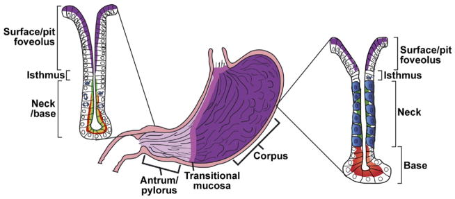
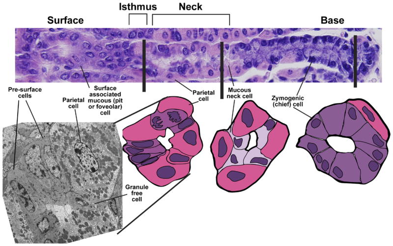
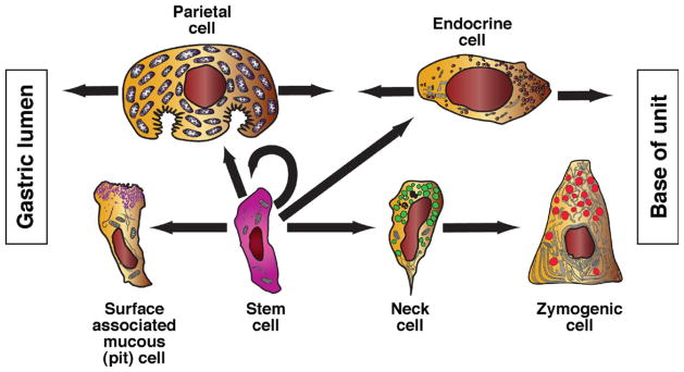
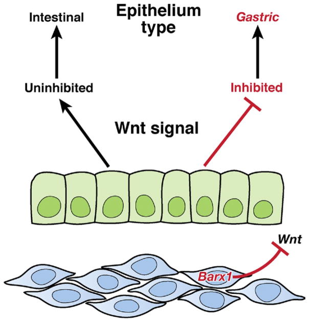
Advances in our understanding of stem cells in the gastrointestinal tract include the identification of molecular markers of stem and early progenitor cells in the small intestine. Although gastric epithelial stem cells have been localized, little is known about their molecular biology. Recent reports describe the use of inducible Cre recombinase activity to indelibly label candidate stem cells and their progeny in the distal stomach, (ie, the antrum and pylorus). No such lineage labeling of epithelial stem cells has been reported in the gastric body (corpus). Among stem cells in the alimentary canal, those of the adult corpus are unique in that they lie close to the lumen and increase proliferation following loss of a single mature progeny lineage, the acid-secreting parietal cell. They are also unique in that they neither depend on Wnt signaling nor express the surface marker Lgr5. Because pathogenesis of gastric adenocarcinoma has been associated with abnormal patterns of gastric differentiation and with chronic tissue injury, there has been much research on the response of stomach epithelial stem cells to inflammation. Chronic inflammation, as induced by infection with Helicobacter pylori, affects differentiation and promotes metaplasias. Several studies have identified cellular and molecular mechanisms in spasmolytic polypeptide-expressing (pseudopyloric) metaplasia. Researchers have also begun to identify signaling pathways and events that take place during embryonic development that eventually establish the adult stem cells to maintain the specific features and functions of the stomach mucosa. We review the cytologic, molecular, functional, and developmental properties of gastric epithelial stem cells.


Conflict of interest statement

Conflicts of interest

The authors disclose no conflicts.

Figures

Figure 1
Figure 1
Typical anatomy and histology of a mammalian stomach. There are a number of variations in mammalian gastric anatomy. For example, mice have a forestomach with keratinized squamous epithelium, whereas humans have a pronounced cardiac region with simpler mucous glands that mark the transition region between the esophagus and corpus. However, the most prominent regions in most mammals are a proximal corpus, encompassing most of the stomach volume, and a distal antrum or pylorus. The corpus epithelium is organized into repeating gastric units that are invaginations from the surface and contain multiple cell lineages in 4 distinct zones. In the diagram, acid-secreting parietal cells are blue, digestive enzyme secreting zymogenic (chief) cells are red, mucous neck cells are green, and the mucus-secreting pit cells nearest the surface are purple. In the antrum, the gastric units are simpler, with few parietal or zymogenic cells. Antral units contain 2 distinct types of mucous cells: those lining the surface (purple) are similar to the surface cells of the corpus, and those nearer to the base have properties intermediate between zymogenic cells and mucous neck cells of the corpus (red-yellow). The interfaces between esophagus and corpus and between corpus and antrum are not abrupt but marked by transitional mucosae. Endocrine cells (not depicted) are also present throughout the corpus and antrum epithelium.
Figure 2
Figure 2
Microanatomy of a corpus gastric unit. (Top) A typical mouse gastric unit, stained by H&E, with the gastric lumen to the left and musculature to the right. Below that are transverse sections through the isthmus (note the dividing cell), neck, and base. Cartoons traced from actual H&E tissue sections are shown. Note the close apposition of progenitor cells in the isthmus and mucous neck cells in the neck with mature parietal cells. (Left) A transverse transmission electron micrograph of a section through the isthmus. A granule-free (presumptive gastric epithelial stem) cell is notable for its high nuclear-cytoplasmic ratio and absence of distinguishing granules. Presurface cells, the precursors to surface mucous (pit or foveolar) cells, are distinguished by early granules that are characteristic of this lineage. Again, mature parietal cells are prominent in the isthmus.
Figure 3
Figure 3
Origins of principal corpus epithelial lineages. The self-renewing stem cell gives rise to each of the principal epithelial lineages of the corpus. There is ultrastructural evidence for the transient intermediates for each lineage (eg, presurface cells depicted in Figure 2); however, available evidence indicates greater complexity in the zymogenic lineage, which arises from a long-lived (≥1 week in mice) intermediate, the mucous neck cell, with its own distinct ultrastructure and probable function.
Figure 4
Figure 4
Approaches to investigate stem cell biology. A stem cell (pink) can be characterized by understanding its niche (location), meaning the cells that surround it and affect its activity. Stem cells can also be characterized by identification of molecular markers, which allow their distinction and isolation (allocation) from differentiated cells. Finally, stem cells can be assayed for functional activity based on their ability to regenerate all the normal lineages of a specific tissue (relocation). Ideally, relocation experiments are performed in vivo (eg, serial transfer of hematopoietic stem cells to irradiated recipients). However, tissue culture approaches have also been useful for isolating and demonstrating the properties of stem cells. In several tissues, including the gastric antrum, single cells have been shown to function as stem cells. However, isolation procedures in all tissues only enrich the fraction of single cells that have stem cell capacity; pure populations of single cells, each with complete stem cell activity, have not yet been isolated.
Figure 5
Figure 5
Cellular mechanisms of SPEM. Chronic inflammation of the corpus in mammals leads to characteristic changes in differentiation in the gastric unit. Parietal cells are lost (atrophy), and the zymogenic chief cell lineage is reprogrammed so that genes that are normally expressed only in mucous neck cells, such as spasmolytic polypeptide/TFF2 (shown in green), are expressed at high levels in cells at the base. The zymogenic cell–specific transcription factor MIST1 (not depicted) is lost, but other zymogenic cell markers (such as pepsinogen C; red) are coexpressed with neck cell markers. Proliferation is increased and occurs more basally in the unit. The pattern of basal proliferation and coexpression of neck and zymogenic cell genes is similar to the histologic pattern in the normal antrum and pylorus, which is why it is called pseudopyloric metaplasia. The most common metaplasia-inducing inflammation is caused by H pylori infection, although autoimmune gastritis (in which autoantibodies target parietal cells) can cause the same metaplasia pattern.
Figure 6
Figure 6
Molecular basis of stomach epithelial specification in embryos. Expression of the homeodomain transcription factor Barx1 is restricted to the developing mesenchyme, which underlies the nascent gastric epithelium. Barx1 regulates transcription of many factors, including the secreted inhibitors of Wnt signaling that repress the canonical Wnt pathway in the overlying endoderm. This repression promotes stomach epithelial differentiation at the expense of intestinal differentiation, which would occur in the absence of Barx1-induced Wnt blockade.

References

    1. Suzuki S, Tsuyama S, Murata F. Cells intermediate between mucous neck cells and chief cells in rat stomach. Cell Tissue Res. 1983;233:475–484. - PubMed
    1. Thompson M, Fleming KA, Evans DJ, et al. Gastric endocrine cells share a clonal origin with other gut cell lineages. Development. 1990;110:477–481. - PubMed
    1. Mills JC, Syder AJ, Hong CV, et al. A molecular profile of the mouse gastric parietal cell with and without exposure to Helicobacter pylori. Proc Natl Acad Sci U S A. 2001;98:13687–13692. - PMC - PubMed
    1. Mills JC, Andersson N, Stappenbeck TS, et al. Molecular characterization of mouse gastric zymogenic cells. J Biol Chem. 2003;278:46138–46145. - PubMed
    1. Mills JC, Andersson N, Hong CV, et al. Molecular characterization of mouse gastric epithelial progenitor cells. Proc Natl Acad Sci U S A. 2002;99:14819–14824. - PMC - PubMed