Comparative Study
. 2011 Mar;41(3):661-8.
doi: 10.1016/j.nbd.2010.11.017. Epub 2010 Dec 8.

Cisplatin induced mitochondrial DNA damage in dorsal root ganglion neurons

Affiliations
Comparative Study

Cisplatin induced mitochondrial DNA damage in dorsal root ganglion neurons

Jewel L Podratz et al. Neurobiol Dis. 2011 Mar.

Abstract

Cisplatin is a platinum-based chemotherapeutic agent that induces peripheral neuropathy in 30% of patients. Peripheral neuropathy is the dose limiting side effect, which has no preventative therapy. We have previously shown that cisplatin induces apoptosis in dorsal root ganglion (DRG) sensory neurons by covalently binding to nuclear DNA (nDNA), resulting in DNA damage, subsequent p53 activation and Bax-mediated apoptosis via the mitochondria. We now demonstrate that cisplatin also directly binds to mitochondrial DNA (mtDNA) with the same binding affinity as nDNA. Cisplatin binds 1 platinum molecule per 2166 mtDNA base pairs and 1 platinum molecule per 3800 nDNA base pairs. Furthermore, cisplatin treatment inhibits mtDNA replication as detected by 5-bromo-2'-deoxy-uridine (BrdU) incorporation and inhibits transcription of mitochondrial genes. The relative reduction in mtDNA transcription is directly related to the distance the gene is located from the transcription initiation point, which implies that randomly formed platinum adducts block transcription. Cisplatin treated DRG neurons exhibit mitochondrial vacuolization and degradation in vitro and in vivo. Taken together, this data suggests that direct mtDNA damage may provide a novel, distinct mechanism for cisplatin-induced neurotoxicity separate from the established nDNA damage pathway.


Figures

Figure 1
Figure 1
DRG culture system used to study cisplatin-mtDNA interactions. To inhibit cisplatin-induced apoptosis, we used 100ng/ml NGF to inhibit Bax translocation and cell cycle changes in combination with the pan-caspase inhibitor zVAD-FMK. Cell survival was determined in cultured DRG neurons treated with or without 2μg/ml cisplatin, 10 or 100ng/ml NGF, and with or without 75μM zVAD-FMK. Survival in DRG neurons treated with cisplatin, 100ng/ml NGF and zVAD-FMK was 94% at 48h, 88% at 72h, 84% at 96h and 75% at144h (black arrow, n=3) and was not significantly different from control DRG neuron survival. Addition of 100ng/ml NGF and zVAD-FMK did however, significantly increase cell survival in cisplatin treated DRG neurons (p<0.001).
Figure 2
Figure 2
Cisplatin binds mtDNA and the resulting adducts can be quantitated. (A) A PCR assay using long PCR amplification of a 13.4 kb fragment of the mitochondrial genome was developed to quantitate the number of platinum-mtDNA adducts. Formation of a cisplatin-DNA adduct on mtDNA blocks PCR amplification reducing the amount of PCR product in the assay. (B) Standard curve using rat liver mtDNA exposed to cisplatin in vitro with PCR products and platinum-DNA binding measured in parallel. PCR samples were measured against the linear portion of the curve, 1Pt molecule per 300bp–6000bp (r2=0.9264). (C) Ethidium bromide gel detection of mtDNA PCR products from cultured DRG neurons treated with 2μg/ml cisplatin, 100ng/ml NGF and 75μM zVADFMK for 0, 48 and 144 hours. (D) Ethidium bromide gel quantitation of mtDNA PCR product from cisplatin treated DNA was expressed as a percent of mtDNA PCR product from non-treated DNA (time 0) Cisplatin induced a significant decrease in the 13.4kb mtDNA PCR product at both 48 and 144 hours (***-p<0.001, n=3) and no significant change with the 235bp mtDNA PCR product (n=3). (E) Calculation of cisplatin adducts compared to the standard curve.
Figure 3
Figure 3
BrdU incorporation decreased in cisplatin-treated DRG neurons. (A) Co-localization of the BrdU incorporation into mtDNA (green) and a mitochondrial targeted plasmid, expressing DS-Red (red). (B) Representative images of BrdU incorporation into DRG neurons treated with cisplatin at 0 and 48 hours (100× magnification). BrdU incorporation observed at time 0 was decreased substantially by 48 hours cisplatin treatment. No BrdU incorporation is observed in the nucleus. (C) DRG neurons were exposed to 2μg/ml cisplatin for 0, 48, 72, 96, 144 and 168 hours in the presence of 100ng/ml NGF and 75μM zVAD-FMK. Both the intensity and area of BrdU fluorescence within the cytoplasm was measured. Cisplatin induced a significant decrease in BrdU incorporation at 48, 144 and 168 hours (***-p<0.001, n=38–77) and a less significant at 72 and 96 hours (*-p<0.05, ns, n=59–64). There appeared to be a recovery of BrdU incorporation at 72 and 96 hours, however, BrdU incorporation did not return to control levels.
Figure 4
Figure 4
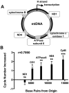
Cisplatin inhibited transcription of mitochondrial genes. (A) Primers were designed to amplify genes transcribed at various locations along the entire mitochondrial genome. (B) DRG neurons were exposed to 2μg/ml cisplatin, 100ng/ml NGF and 75μM zVAD-FMK for 144 hours, harvested, RNA isolated and real time RT-PCR performed. The number of cycles needed to cross the PCR threshold was compared to control cDNA. Changes in PCR cycles increased the farther away the gene was located from the transcription origin. The relationship of the cycle number to the distance from the transcription origin was linear (r2=0.7990). Cycle number had a slight decrease of 0.27 cycles for ND1 (ns, n=5) and an increase of 1.21 cycles for CO1 (*-p<0.05, n=5), 1.48 cycles for ATPase 6 (**-p<0.01, n=5), 1.68 cycles for ND4 (**-p<0.01, n=5) and 2.21 cycles for Cyt b (***-p<0.001, n=5).
Figure 5
Figure 5
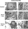
Cisplatin induced mitochondrial degradation and vacuolization in cultured DRG neurons. TEM of intact (a, arrows) normal mitochondria and (b, *) nuclear membrane in control DRG neurons. DRG neurons treated with 2μg/ml cisplatin 100ng/ml NGF and 75μM zVAD-FMK showed degradation of mitochondria (c) at 48 hours that increased (e) at 144 hours. The (d, f) nuclear membrane, however, remained intact.
Figure 6
Figure 6
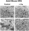
Adult mice were treated with vehicle (saline) or cisplatin (23mg/kg, cumulative). DRG were harvested and examined by TEM. Control DRG treated with saline showed normal mitochondria (arrows) and intact nuclear membrane (*). Cisplatin treated DRG showed mitochondrial degradation (arrows) in the presence of intact nuclear membrane (*).

References

    1. Asin-Cayuela J, Gustafsson CM. Mitochondrial transcription and its regulation in mammalian cells. Trends Biochem Sci. 2007;32:111–7. - PubMed
    1. Bottone MG, et al. Cell proliferation, apoptosis and mitochondrial damage in rat B50 neuronal cells after cisplatin treatment. Cell Prolif. 2008;41:506–20. - PMC - PubMed
    1. Cavaletti G. Peripheral neurotoxicity of platinum-based chemotherapy. Nat Rev Cancer. 2008;8 1p following 71; author reply 1p following 71. - PubMed
    1. Clayton DA. Replication and transcription of vertebrate mitochondrial DNA. Annu Rev Cell Biol. 1991;7:453–78. - PubMed
    1. Conti AM, et al. Inhibition of axonal growth from sensory neurons by excess nerve growth factor. Ann Neurol. 1997;42:838–846. - PubMed

Publication types