. 2010 Dec 10;40(5):749-61.
doi: 10.1016/j.molcel.2010.09.029.

Taf1 regulates Pax3 protein by monoubiquitination in skeletal muscle progenitors

Affiliations

Taf1 regulates Pax3 protein by monoubiquitination in skeletal muscle progenitors

Stéphane C Boutet et al. Mol Cell. .

Abstract

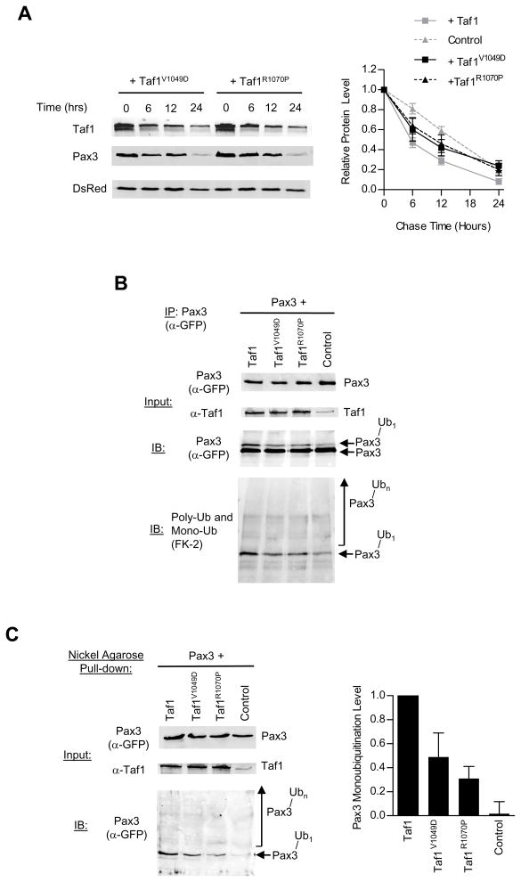
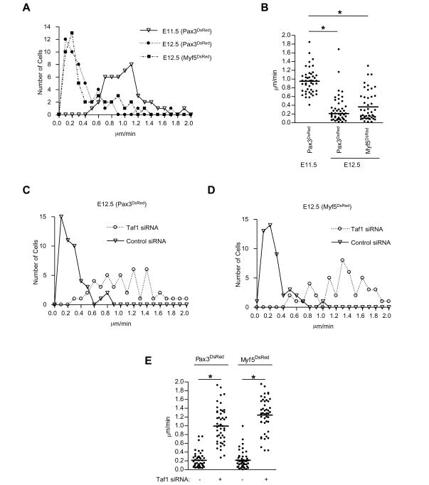
Pax3 plays critical roles during developmental and postnatal myogenesis. We have previously shown that levels of Pax3 protein are regulated by monoubiquitination and proteasomal degradation during postnatal myogenesis, but none of the key regulators of the monoubiquitination process were known. Here we show that Pax3 monoubiquitination is mediated by the ubiquitin-activating/conjugating activity of Taf1, a component of the core transcriptional machinery that was recently reported to be downregulated during myogenic differentiation. We show that Taf1 binds directly to Pax3 and overexpression of Taf1 increases the level of monoubiquitinated Pax3 and its degradation by the proteasome. A decrease of Taf1 results in a decrease in Pax3 monoubiquitination, an increase in the levels of Pax3 protein, and a concomitant increase in Pax3-mediated inhibition of myogenic differentiation and myoblast migration. These results suggest that Taf1 regulates Pax3 protein levels through its ability to mediate monoubiquitination, revealing a critical interaction between two proteins that are involved in distinct aspects of myogenic differentiation. Finally, these results suggest that the components of the core transcriptional are integrally involved in the process of myogenic differentiation, acting as nodal regulators of the differentiation program.


Conflict of interest statement

Competing Interests statement The authors declare that they have no competing financial interests.

Figures

Figure 1
Figure 1. Taf1 regulates Pax3 protein levels and binds to Pax3
(A) Western blot analysis of the expression level of Taf1 and Pax3 after treatment with Taf1 siRNA and before induction of differentiation. Primary myoblasts were transfected with Taf1 or control siRNA (60 nM each). Cells were harvested and analyzed by immunoblots with anti-Taf1, anti-Pax3, and anti-GAPDH antibodies. (B) Quantitative analysis of Taf1 and Pax3 mRNA levels in satellite cells from day 0 through day 5 of activation. Each is normalized to GAPDH; the level at the peak of expression was arbitrarily set at 1. (C) Western blot analysis of Taf1 and Pax3 levels at day 0 through day 5 of activation. (D) Analysis of interaction between Taf1 and exogenous Pax3. C2C12 myoblasts transfected with Pax3-GFP or Control (empty vector) constructs were treated for 3 hours with MG132 and lysed. Proteins were immunoprecipitated using an anti-GFP antibody and analyzed by immunoblotting using anti-Taf1 and anti-Taf4 antibodies. (E) Analysis of interaction between exogenous Pax3 and Taf1 in cells. C2C12 myoblasts were transfected with HA-tagged hTaf1 and either Pax3-GFP or GFP expression constructs, treated for 3 hours with MG132, and lysed. Proteins were immunoprecipitated using an anti-HA antibody and analyzed by immunoblotting using anti-HA and anti-GFP antibodies. (F) Analysis of endogenous Pax3 and Taf1 interaction. Primary myoblasts were treated for 24 hours with MG132 (5 μM) to allow accumulation of ubiquitinated Pax3 and lysed. Proteins were immunoprecipitated using an anti-Taf1 antibody and analyzed by immunoblotting using an anti-Pax3 antibody. The position of the band corresponds to the molecular weight of monoubiquitinated Pax3. (G) Analysis of direct interactions between Pax3 and Taf1. Purified HA-tagged hTaf1 was immunoprecipitated using an anti-HA affinity matrix and mixed with purified recombinant Pax3 proteins. Pax3 proteins were analyzed by immunoblotting using an anti-Pax3 antibody. (See also Figure S1)
Figure 2
Figure 2. Taf1 is both sufficient and necessary for Pax3 monoubiquitination
(A) Analysis of Pax3 protein stability by pulse-chase experiments in C2C12 myoblasts transfected with constructs expressing Pax3 and DsRed, and with either a control vector or a Taf1-expression vector. The cells were treated with cycloheximide for the indicated times. In each case, the level of DsRed was used as an internal control. Protein levels were assessed by immunoblot analysis (left panel). Quantitative analysis of replicate experiments (right panel) shows increased degradation of Pax3 in Taf1-expressing cells. (B) Analysis of Pax3 ubiquitination by Taf1 in a cell-free ubiquitination assay. Reactions containing purified ubiquitin, HA-hTaf1 and Pax3 in the presence or absence of ATP were analyzed by immunoblots with anti-Pax3, anti-Taf1 and anti-ubiquitin antibodies. Arrows show the positions of Pax3 and monoubiquitinated Pax3 (Pax3-Ub1). (C) Western blot analysis of Pax3 levels in myoblasts with either normal or reduced levels of Taf1. Primary myoblasts were transfected with Taf1 siRNA or control siRNA at the indicated concentration. Pax3 and Taf1 protein levels were assessed by immunoblot analysis and, in each case, the level of GAPDH was used as an internal control. Representative blots are shown above, and quantitation of replicate experiments is shown below. (D) Accumulation of non-ubiquitinated Pax3 after Taf1 downregulation. Primary myoblasts were treated with 60 nM Taf1 or Control siRNA. After 24 hours, cells were incubated with MG132 for 3 hours to allow accumulation of monoubiquitinated Pax3. Pax3 levels were analyzed by immunoblots with an anti-Pax3 antibody. Arrows show the positions of monoubiquitinated and unmodified Pax3. Under the conditions of reduced Taf1, there is a marked accumulation of the non-ubiquitinated form whereas in the absence of Taf1 siRNA, nearly all of the protein is monoubiquitinated (and detectable only because of the addition of MG132 to the cultures). (E) Quantitative analysis of replicate immunoblot studies as shown in panel (D) showing the ratio of non-ubiquitinated Pax3 to total Pax3. The relative level of Taf1 protein (grey squares) in cells treated with Taf1 or control siRNA oligonucleotides is superimposed (scale to the right). The ratio of non-ubiquitinated Pax3/total Pax3 increased markedly when Taf1 was downregulated, even though there was a concomitant increase in monoubiquitinated Pax3 because of the marked increase of the non-ubiquitinated form. The value with the control siRNA treatment was arbitrarily set at 1.0. All data are normalized to GAPDH.
Figure 3
Figure 3. Mutation in the Taf1 UBAC domain reduces Pax3 monoubiquitination and degradation
(A) Analysis of Pax3 protein stability by pulse-chase experiments in C2C12 myoblasts transfected with constructs expressing Pax3 and DsRed, and with constructs expressing either Taf1V1046D or Taf1R1070P. The cells were treated with cycloheximide for the indicated times. In each case, the level of DsRed was used as an internal control. Protein levels were assessed by immunoblot analysis (left panel). Quantitative analysis of replicate experiments (right panel) shows levels of Pax3 in mutant Taf1-expressing cells that are intermediate between those in control cells and those in Taf1-expressing cells. The data from Figure 2A showing Pax3 levels in control cells or cells expressing wild-type Taf1 are presented in light grey as reference (the studies were done concurrently). (B) Analysis of the level of ubiquitination of Pax3 in the presence of overexpressed wild-type and mutant Taf1. C2C12 myoblasts transfected with Pax3-GFP and either Taf1, Taf1V1046D, Taf1R1070P, or control expression constructs were treated for 3 hours with MG132 to allow accumulation of ubiquitinated Pax3 and lysed. Proteins were denatured by boiling and analyzed by immunoblotting using an anti-GFP antibody (for Pax3 proteins) or FK-2 antibody (recognizes both polyubiquitinated and monoubiquitinated proteins) antibodies. Arrows show the positions of Pax3, monoubiquitinated Pax3 (Pax3-Ub1), and where any polyubiquitinated Pax3 (Pax3-Ubn) would migrate if present. (C) Analysis of the level of ubiquitination of Pax3 in presence of overexpressed wild-type or mutant Taf1 in vivo. C2C12 myoblasts transfected with Pax3-GFP, His6-tagged ubiquitin and either Taf1, Taf1V1046D, Taf1R1070P, or control expression constructs were treated for 3 hours with MG132 to allow accumulation of ubiquitinated Pax3 and lysed. His6-tagged ubiquitin conjugates were purified on Nickel-agarose gels and levels of Pax3 proteins were analyzed by immunoblotting with an anti-GFP antibody. Again, arrows show the positions of monoubiquitinated Pax3 (Pax3-Ub1) and where any polyubiquitinated Pax3 (Pax3-Ubn) would migrate if present. Monoubiquitination levels (shown quantitatively on the right) were determined as the ratio of monoubiquitinated Pax3 protein to Pax3 protein input and then normalized to the value obtained for wild-type Taf1. (See also Figure S2)
Figure 4
Figure 4. Functional regulation of myogenesis by Taf1
(A) Western blot and quantitative analysis of the level of expression of myogenic differentiation markers in cells with normal or reduced Taf1. Primary myoblasts were transfected with Taf1 or Control siRNA (60 nM each) and switched to differentiation medium 24 hours after transfection for an additional 24 hours. Cells were then harvested and analyzed by immunoblots with anti-Myogenin (MyoG), anti-Sarcomeric α-Actinin, and anti-Myosin Heavy Chain (MyHC) antibodies. Each is normalized to GAPDH. The levels in Control siRNA treated cells were arbitrarily set to 1. Representative blots are to the left, and quantitation of replicate experiments is shown to the right. (B) Enhancement of differentiation by Taf1 regulation is mediated by Pax3. Western blot and quantitative analysis of the level of expression of myogenic differentiation markers in myogenic progenitors with reduced Taf1 alone or with reduced Pax3. Primary myoblasts were transfected with Taf1 siRNA and either Pax3 or control siRNA (60 nM each) and switched to differentiation medium 24 hours after transfection for an additional 24 hours. Cells were then harvested and analyzed by immunoblots with anti-Myogenin (MyoG), anti-Sarcomeric α-Actinin, and anti-Myosin Heavy Chain (MyHC) antibodies. Each is normalized to GAPDH protein level and the levels in Taf1 and Pax3 siRNA treated cells were arbitrarily set to 1. Representative blots are shown to the left, and quantitation of replicate experiments is shown to the right. (See also Figure S3)
Figure 5
Figure 5. Distribution of cell motility of primary myoblasts as regulated by Pax3 and Taf1
(A) Primary myoblasts were transfected with Taf1 siRNA, Pax3 siRNA, control siRNA (60 nM each), or lipofectamine alone. Twenty-four hours after transfection, cell motility was assessed by recording cell positions every 5 minutes for 3 hours. Forty-six cells over three independent experiments were used for analysis. Each point represents the number cells in the population that exhibited the average motility indicated on the abscissa. Cell populations treated with Taf1 siRNA were significantly more motile than cell populations treated with control siRNA, Pax3 siRNA or lipofectamine alone. (B) Scatter plot representation of cell motility by time-lapse microscopy shown in panel (A). Taf1 siRNA-treated primary myoblasts were significantly faster than control siRNA treated myoblasts (*p<0.0001). Median is indicated as a horizontal bar. (C) Enhancement of migration by Taf1 regulation is mediated by Pax3. Primary myoblasts were transfected with Taf1 siRNA and either Pax3 or control siRNA (60 nM each) or lipofectamine alone. Twenty-four hours after transfection, cell motility was recorded for 3 hours every 5 minutes. Analysis was identical to that in panel (A). The knockdown of Pax3 abrogated the enhanced motility by Taf1 knockdown. (D) Scatter plot representation of cell motility by time-lapse microscopy shown in panel (C). Taf1 siRNA treated primary myoblasts were significantly faster than Taf1 and Pax3 siRNA treated myoblasts (*p<0.0001). Median is indicated as a horizontal bar. (See also Figure S4)
Figure 6
Figure 6. Pax3 and Taf1 expression in E11.5 and E12.5 embryonic myogenic progenitors
(A) Expression of Pax3 and Taf1 in Pax3DsRed and Myf5DsRed sorted myogenic cells at E11.5 and E12.5. Cells were harvested and analyzed by immunoblots with anti-Pax3, anti-Taf1 and anti-GAPDH antibodies. (B) Quantitative analysis of replicate immunoblot studies shown in panel (A). Protein levels were normalized to GAPDH and then to the values at E11.5. (C) Quantitative analysis by qRT-PCR of Pax3 and Taf1 mRNA levels in progenitor cells isolated from embryos at E11.5 (Pax3DsRed) and at E12.5 (Pax3DsRed and Myf5DsRed). Transcript levels were normalized to GAPDH and then to the values at E11.5. (D) Analysis of Pax3 protein levels in embryonic progenitors after treatment with MG132. E11.5 limbs were harvested and treated for 24 hours with DMSO or MG132 (10 μM). After treatment, limbs were harvested and lysed. Proteins were detected by Western blotting with anti-Pax3, anti-Taf1 and anti-GAPDH antibodies. Under these conditions, nearly all of the protein is in the monoubiquitinated form. (See also Figure S5)
Figure 7
Figure 7. Analysis of the motility of embryonic myogenic progenitors by time lapse microscopy
(A) The motility of myogenic cells isolated from limbs of embryos at E11.5 (Pax3DsRed) and E12.5 (Pax3DsRed or Myf5DsRed) was determined by recording cell positions every 5 minutes for 3 hours. (B) These scatter plots present the distribution of motilities for the graphs shown in panel (A). Myogenic progenitors at E11.5 were significantly more motile than those at E12.5 (*p<0.0001). Median is indicated as a horizontal bar. (C,D) Sorted cells at E12.5 either from Pax3DsRed (C) or Myf5DsRed (D) mice were plated on laminin/collagen, transfected with Taf1 or control siRNA (60 nM each), and assessed for motility by recording cell position every 5 minutes for 3 hours. Cells treated with Taf1 siRNA were significantly more motile than cells treated with control siRNA. (E) Scatter plot representation of data in panels (C) and (D) of myogenic progenitors isolated from embryos at E12.5 (Pax3DsRed or Myf5DsRed) transfected with Taf1 or control siRNA. Taf1 siRNA treated myoblasts were significantly more motile than control siRNA treated myoblasts (*p<0.0001). Median is indicated as a horizontal bar. (See also Figure S6)

References

    1. Bajard L, Relaix F, Lagha M, Rocancourt D, Daubas P, Buckingham ME. A novel genetic hierarchy functions during hypaxial myogenesis: Pax3 directly activates Myf5 in muscle progenitor cells in the limb. Genes Dev. 2006;20:2450–2464. - PMC - PubMed
    1. Belz T, Pham AD, Beisel C, Anders N, Bogin J, Kwozynski S, Sauer F. In vitro assays to study protein ubiquitination in transcription. Methods. 2002;26:233–244. - PubMed
    1. Bertolotti A, Lutz Y, Heard DJ, Chambon P, Tora L. hTAF(II)68, a novel RNA/ssDNA-binding protein with homology to the pro-oncoproteins TLS/FUS and EWS is associated with both TFIID and RNA polymerase II. EMBO J. 1996;15:5022–5031. - PMC - PubMed
    1. Biressi S, Molinaro M, Cossu G. Cellular heterogeneity during vertebrate skeletal muscle development. Dev Biol. 2007;308:281–293. - PubMed
    1. Bladt F, Riethmacher D, Isenmann S, Aguzzi A, Birchmeier C. Essential role for the c-met receptor in the migration of myogenic precursor cells into the limb bud. Nature. 1995;376:768–771. - PubMed

Publication types

MeSH terms

Substances

LinkOut - more resources