. 2010 Dec 14;33(6):942-54.
doi: 10.1016/j.immuni.2010.11.022. Epub 2010 Dec 9.

Reprogrammed foxp3(+) regulatory T cells provide essential help to support cross-presentation and CD8(+) T cell priming in naive mice

Affiliations

Reprogrammed foxp3(+) regulatory T cells provide essential help to support cross-presentation and CD8(+) T cell priming in naive mice

Madhav D Sharma et al. Immunity. .

Abstract

Foxp3(+) regulatory T (Treg) cells can undergo reprogramming into a phenotype expressing proinflammatory cytokines. However, the biologic significance of this conversion remains unclear. We show that large numbers of Treg cells undergo rapid reprogramming into activated T helper cells after vaccination with antigen plus Toll-like receptor 9 (TLR-9) ligand. Helper activity from converted Treg cells proved essential during initial priming of CD8(+) T cells to a new cross-presented antigen. Help from Treg cells was dependent on CD40L, and (unlike help from conventional non-Treg CD4(+) cells) did not require preactivation or prior exposure to antigen. In hosts with established tumors, Treg cell reprogramming was suppressed by tumor-induced indoleamine 2,3-dioxygenase (IDO) and vaccination failed because of lack of help. Treg cell reprogramming, vaccine efficacy, and antitumor CD8(+) T cell responses were restored by pharmacologic inhibition of IDO. Reprogrammed Treg cells can thus participate as previously unrecognized drivers of certain early CD8(+) T cell responses.


Figures
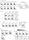
Figure 1
Figure 1. Foxp3+ Treg cells undergo phenotypic reprogramming following vaccination
(A) Foxp3GFP mice received adoptive transfer of OT-I cells and immunization with OVA+CpG+IFA vaccine. At different time points, draining LNs were stained for phospho-STAT5 and surface CD69; graphs show the percentage of gated CD4+ Treg cells (GFP+) and non-Treg cells (GFP) expressing the antigens at each time-point. (B) Foxp3GFP mice received OT-I cells and were immunized as above, with or without CpG in the vaccine. Draining LN cells were harvested 4 days after vaccination, incubated for 4 hrs with low-dose PMA+ionomycin in the presence of brefeldin A, then stained for intracellular cytokines. (C) Foxp3GFP mice received OT-I cells and immunization as above, and draining LNs were stained for CD40L expression at different times (staining was directly ex vivo, without PMA treatment). Graph shows pooled data from 3–5 experiments at each time-point (bars show SD). (D) F1(Foxp3-GFP-cre × ROSA26-YFP) mice received OT-I cells and immunization as above, and draining LNs were analyzed on day 4. GFP and YFP are both seen in the FL1 channel. (E) Foxp3GFP mice received CFSE-labeled OT-I cells (Thy1.1 congenic), followed by OVA vaccine containing IFA plus graded amounts of CpG. CD4+ cells were analyzed for surface CD40L; OT-I cells (gated Thy1.1+) were analyzed for CFSE dye-dilution and intracellular granzyme B. In each panel, data are representative of 3–5 experiments (17 experiments for panel B). See also Supplemental Figure S1.
Figure 2
Figure 2. Reprogrammed Treg cells are functionally required to support early CD8+ T cell response to a cross-presented vaccine antigen
(A–C) T cell-deficient Tcra/ host mice received adoptive transfer of sorted Treg cells or CD4+ non-Treg cells as indicated. Mice were rested for 1–7 days, then received CFSE-labeled OT-I cells and immunization with OVA+CpG+IFA. Responses were analyzed in draining LNs after 4 days. (A) Foxp3GFP Treg cells or non-Treg cells (CD4+GFP) were transferred; controls received no CD4+ cells. Comparison of OT-I cell responses in draining LN. (B) Treg cells (CD4+CD25+) were sorted from WT B6 or Cd40lg/ mice and transferred into Tcra/ hosts (controls received no CD4+ cells), followed by adoptive transfer of OT-I cells and vaccination with OVA+CpG+IFA. Draining LNs were analyzed for DCs (CD11c+) and OT-I cells (CD8+). (C) Tcra/ mice received Foxp3GFP Treg cells or no Treg cells. A third group received no Treg cells but were treated with anti-CD40 (clone FGK45, 250 ug i.p. on the day of vaccination and 100 ug i.p. 2 days later). Plots show CFSE-labels OT-I cells in vaccine-draining LNs on day 4. (D) CD40L-deficient host model: Cd40lg/ mice received adoptive transfer of CD40L-sufficient Foxp3GFP Treg cells (2–4 × 10⁵ CD4+GFP+) or non-Treg cells (1 × 10⁶ CD4+GFP), both congenically marked with Thy1.1. All mice received CFSE-labeled OT-I cells and immunization as above, and response of OT-I cells and transferred CD4+ cells analyzed on day 4. In each panel, data are representative of 3–6 experiments. See also Supplemental Figure S2.
Figure 3
Figure 3. Progressive unresponsiveness to vaccination induced by established tumors
C57BL/6 mice were implanted with 1 × 10⁶ B16F10 tumor cells (control mice received no tumor). After 3–7 days of tumor growth, mice received adoptive transfer of CFSE-labeled resting pmel-1 cells (sorted CD8+) and gp100+CpG+IFA vaccine. Four days later, tumor-draining LNs were analyzed (or vaccine-draining LN in mice without tumors). Representative of a total of 9 experiments on day 7, and 3 experiments each on other days.
Figure 4
Figure 4. Treg cell reprogramming in tumor-bearing hosts is antagonized by IDO, and restored by pharmacologic IDO-inhibitor
(A) Foxp3GFP host mice were implanted with B16F10 tumors. On day 7 of tumor growth, mice received adoptive transfer of resting pmel-1 cells (sorted CD8+), with or without gp100+CpG+IFA vaccine. Groups were treated either with continuous 1MT in drinking water (beginning on day 6, one day prior to vaccination) or with vehicle control, as indicated. Four days after vaccination, tumor-draining LNs were harvested, treated with low-dose PMA+ionomycin, and stained as in Figure 1. The lower set of dot-plots show the gated GFP+ (Treg cell) population. (B) One day prior to tumor implantation, Treg cells (CD4+CD25+) were enriched from GCN2-deficient Eif2ak4/ mice or from WT B6 controls, and transferred into congenic Thy1.1+ hosts. B16F10 tumors were then implanted, and 7 days later mice received pmel-1 cells and vaccine as in the previous panel. (None of the mice were treated with 1MT). After 4 days, the transferred Treg cells were analyzed in tumor-draining LNs. (C) Prior to tumor implantation, T cell-deficient Tcra/ hosts received a mixture of CD4+ cells, comprising Thy1.2+ Treg cells from Foxp3GFP donors plus congenic Thy1.1+ non-Treg cells (CD4+GFPThy1.1+). All mice then received B16F10 tumors, and 7 days later were treated with pmel-1 cells and vaccine, with or without oral 1MT as indicated. Four days after vaccination, tumor-draining LNs were analyzed for transferred Treg cell and non-Treg populations, gated separately on Thy1 allotype. Each panel is representative of 3–6 experiments. See also Supplemental Figure S3.
Figure 5
Figure 5. Inhibition of IDO enhances efficacy of anti-tumor vaccination by restoring help from reprogrammed Treg cells
(A) C57BL/6 mice with day 7 established B16F10 tumors received CFSE-labeled pmel-1 cells and vaccination with gp100+CpG+IFA, with or without 1MT in drinking water as shown. Four days later, gated pmel-1 cells (Thy1.1+) were analyzed in tumor-draining LN and disaggregated tumor. Representative of 6 experiments with tumors and 11 experiments with tumor-draining LNs. Tumor size (product of orthogonal diameters) is shown after dissection at necropsy (day 11). * p<.01 by t-test (n=16 tumors in 8 separate experiments) (B) Tcra/ hosts received pre-adoptive transfer of Foxp3GFP Treg cells, or CD4+GFP non-Treg cells, or no CD4+ cells, as indicated. Mice were then implanted with B16F10 tumors, and on day 7 received pmel-1 cells and gp100+CpG+IFA vaccine, with or without 1MT as shown. Four days later, pmel-1 cells were analyzed in tumor-draining LN, and tumor size measured at necropsy. Pooled data from 13 experiments (number of tumors indicated in the bar graph, * p<.05 by ANOVA). (C) Treg cells (CD4+CD25+) were sorted from GCN2-deficient Eif2ak4/ mice or WT control donors and 2 × 10⁵ cells transferred into WT (C57BL/6) hosts. All mice were then implanted with B16F10 tumors, and on day 7 received pmel-1 cells, gp100+CpG+IFA vaccination and 1MT as shown. CFSE-labeled pmel-1 cells were measured in TDLNs on day 11. Representative of 3 experiments; * p<.05 by ANOVA). See also Supplemental Figure S4.
Figure 6
Figure 6. Helper activity of reprogrammed Treg cells in tumor-bearing hosts is mediated via CD40L
(A) C57BL/6 or Cd40lg/ hosts were implanted with B16F10 tumors, then on day 7 received pmel-1 cells and gp100+CpG+IFA vaccine, with or without oral 1MT as indicated. Four days later, DCs (CD11c+) were analyzed in tumor-draining LNs. Control mice received no tumor and no treatment. (B) Tcra/ mice received pre-transfer of a mixed CD4+ population comprising 1 × 10⁶ non-Treg cells (CD4+GFP cells from Foxp3GFP mice) plus 2 × 10⁵ Treg cells (enriched as CD4+CD25+) from either C57BL/6 or Cd40lg/ mice. All mice were implanted with B16F10 tumors, and on day 7 received pmel-1 cells and gp100+CpG+IFA vaccine, with or without 1MT as shown. Four days later, pmel-1 cells were analyzed in tumor-draining LNs, and tumor size measured at necropsy (* p<.05 by ANOVA; n per group shown in the figure). (C) Tcra/ mice received pre-transfer of Treg cells (CD4+CD25+) from either WT or Cd40lg/ mice, as shown. All mice were implanted with B16F10 tumors, and on day 7 received pmel-1 cells and gp100+CpG+IFA vaccine, with or without 1MT as indicated. One group also received activating anti-CD40 (clone FGK45, as in Figure 2C). In each panel, data are representative of 3–5 experiments. See also Supplemental Figure S5.

Comment in

References

    1. Antony PA, Piccirillo CA, Akpinarli A, Finkelstein SE, Speiss PJ, Surman DR, Palmer DC, Chan CC, Klebanoff CA, Overwijk WW, et al. CD8+ T cell immunity against a tumor/self-antigen is augmented by CD4+ T helper cells and hindered by naturally occurring T regulatory cells. J Immunol. 2005;174:2591–2601. - PMC - PubMed
    1. Bennett SR, Carbone FR, Karamalis F, Flavell RA, Miller JF, Heath WR. Help for cytotoxic-T-cell responses is mediated by CD40 signalling. Nature. 1998;393:478–480. - PubMed
    1. Bennett SR, Carbone FR, Karamalis F, Miller JF, Heath WR. Induction of a CD8+ cytotoxic T lymphocyte response by cross-priming requires cognate CD4+ T cell help. J Exp Med. 1997;186:65–70. - PMC - PubMed
    1. Cohn M. How does the immune response get started? Cell Immunol. 2009;254:91–93. - PMC - PubMed
    1. Duarte JH, Zelenay S, Bergman ML, Martins AC, Demengeot J. Natural Treg cells spontaneously differentiate into pathogenic helper cells in lymphopenic conditions. Eur J Immunol. 2009;39:948–955. - PubMed

Publication types

MeSH terms