. 2011 Mar 7;10(3):260-70.
doi: 10.1016/j.dnarep.2010.11.007. Epub 2010 Dec 10.

Toxicity and tolerance mechanisms for azidothymidine, a replication gap-promoting agent, in Escherichia coli

Affiliations

Toxicity and tolerance mechanisms for azidothymidine, a replication gap-promoting agent, in Escherichia coli

Deani L Cooper et al. DNA Repair (Amst). .

Abstract

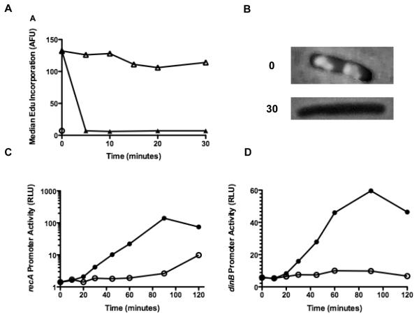
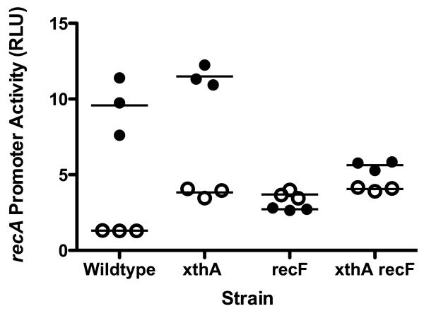
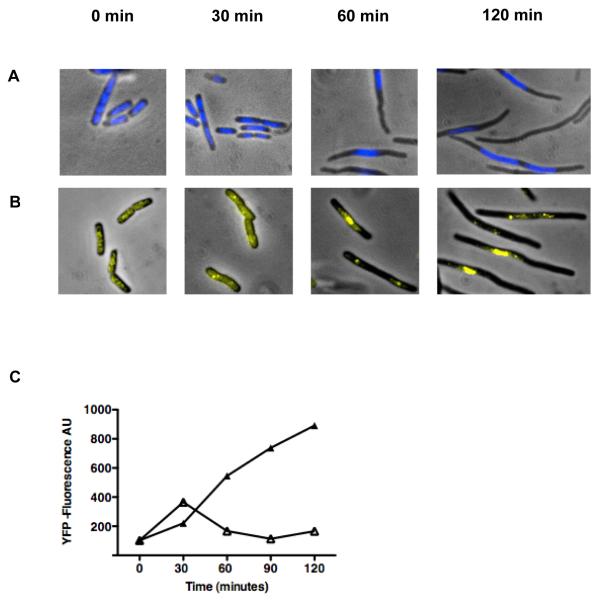
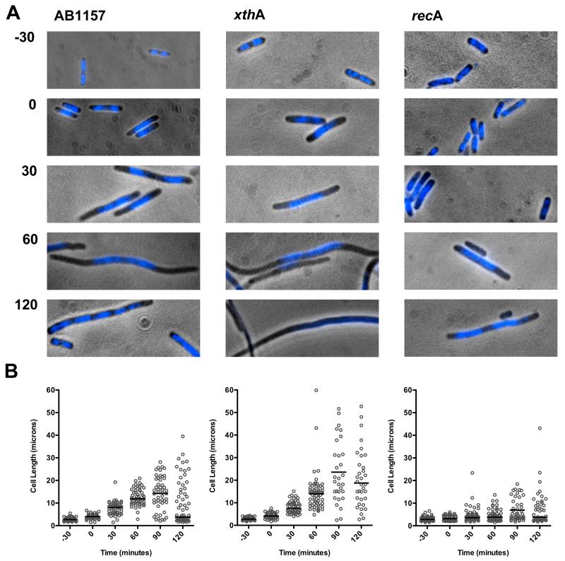
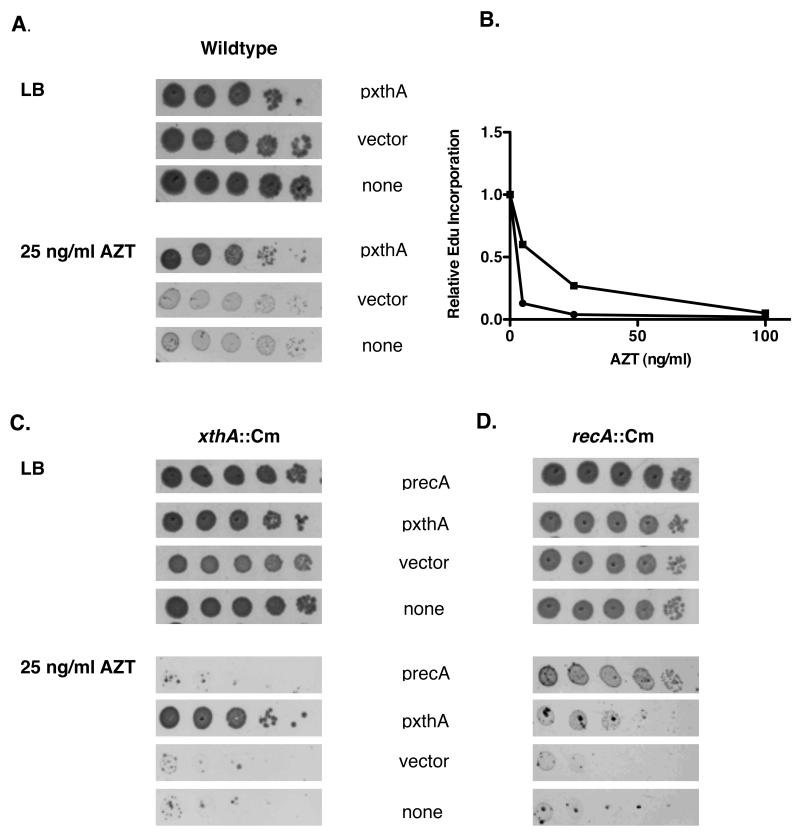
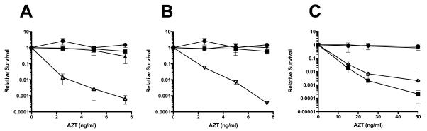
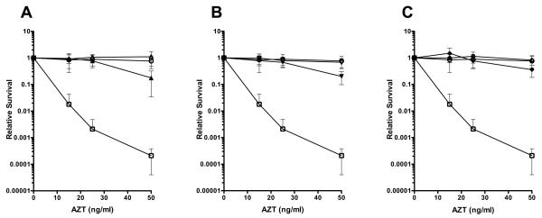
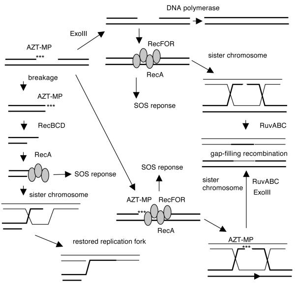
Azidothymidine (AZT, zidovudine) is used to treat HIV-AIDS and prevent maternal transmission to newborns. Because the azido group replaces the 3' OH of thymidine, AZT is believed to act as a chain terminator during reverse transcription of viral RNA into DNA, although other mechanisms of viral inhibition have been suggested. There is evidence that AZT is genotoxic, particularly to the mitochondria. In this study, we use the bacterium Escherichia coli to investigate the mechanism of AZT toxicity and the cellular mechanisms that aid survival. We show that that replication arrests quickly after treatment, accompanied by induction of the SOS DNA damage response. AZT appears to produce single-strand DNA gaps, as evident by RecF-dependent induction of the SOS response and visualization of single-strand DNA binding protein foci within the cell. Some of these gaps must be converted to breaks, since mutants in the RecBCD nuclease, required for recombinational double-strand break repair, are highly sensitive to AZT. Blocks in the late recombination functions, the RuvAB branch migration helicase and RuvC Holliday junction endonuclease, caused extreme AZT sensitivity that could be relieved by mutations in the early recombination functions, such as RecF, suggesting gaps engage in recombination reactions. Finally, our data suggest that the proofreading exonucleases of DNA polymerases play little role in AZT tolerance. Rather, Exonuclease III appears to be the enzyme that removes AZT: xthA mutants are highly AZT-sensitive, with a sustained SOS response, and overproduction of the enzyme protects wild-type cells. Our findings suggest that incorporation of AZT into human nuclear and mitochondrial DNA has the potential to promote genetic instability and toxicity through the production of ssDNA gaps and dsDNA breaks, and predicts that the human Exonuclease III ortholog, APE1, will be important for drug tolerance.


Figures

Figure 1
Figure 1. AZT halts replication and induces SOS
(A) AB1157 cultures were grown to early log phase and then treated with 100 ng/ml AZT or thymidine for the times indicated. At each time point, 2 ml of culture was removed, EdU was added to 40 μg /ml and cultures were incubated for an additional 5 minutes. The cells were then fixed with 90% methanol, labeled with AlexaFluor488 and analyzed as described in the materials and methods. Median fluorescence for 50,000 cells is shown at each time point (⦿ No Edu, △ Thymidine, ▲AZT). (B) Micrograph showing AB1157 cultures labeled with EdU and visualized with Alexa Fluor 488 at 0 and 30 minutes after the addition of 100 ng/ml AZT. (C) AB1157 cultures harboring a plasmid with the luxCDABE operon under control of the recA promoter were grown as in A. At each time point, 0.1 ml of culture was removed and luminescence was quantified using a liquid scintillation counter (⦿ Thymidine, AZT). Values shown are normalized to the A590 of the culture at each time point. (D) As in C, except the plasmid contains the luxCDABE operon under the dinB promoter (⦿ Thymidine, ● AZT).
Figure 2
Figure 2. SOS induction in strains treated with AZT
Strains containing a plasmid with the lux genes under the recA promoter were grown to early log phase and then AZT was added to 150 ng/ml or mock treated. After 40 minutes, culture was removed and luminescence was determined using a liquid scintillation counter. Luminescence was normalized to culture A590 to give relative luciferase units (RLU). (⦿ mock treated cultures, ● treated cultures).
Figure 3
Figure 3. AB1157 cells elongate and SSB foci accumulate after the addition of AZT
(A) Wild type cells stained with DAPI at 0, 30, 60, and 120 minutes after addition of AZT to 100 ng/ml. (B) Wild type cells with a SSB-YPet gene under the natural SSB promoter at 0, 30, 60, and 120 minutes after addition of AZT to 100 ng/ml. (C) Quantification of SSB-YPet accumulation (50,000 cell sample) using flow cytometry (△ Thymidine, ▲ AZT).
Figure 4
Figure 4. Cellular morphology during recovery from AZT treatment
Wild-type, xthA, or recA cultures were grown to early log phase. AZT (100 ng/ml) was then added to the media for 30 minutes after which the cells were collected by centrifugation, washed, and then resuspended in LB. Growth was continued in media without AZT and samples were collected at the times indicated. (A) Micrographs showing cultures stained with DAPI before treatment with AZT (−30), immediately after treatment with AZT for 30 minutes (0), or after AZT was removed from the cultures for 30, 60, or 120 minutes. (B) Lengths of at least 50 cells from 2 slides were measured using Volocity Openlab software. Each individual cell length is represented with a ○ and the median cell length for each population is indicated by the horizontal bar.
Figure 5
Figure 5. Overproduction of Exonuclease III but not RecA mitigates AZT toxicity
(A) Wild type cultures transformed with vector (pBSSK-) or a plasmid over-expressing xthA (in pBSSK-) were grown at 37° to an A590 of approximately 0.4 in LB media with 100 mg/ml Ap. Cultures were then serially diluted with 1X 56/2 media supplemented with 2mM IPTG. An additional 50 μg /ml ampicillin was added to serial dilutions when a plasmid was present. Diluted cultures were plated on either LB plates or LB plates with 25 ng/ml AZT. Plates were then incubated at 37° for approximately 24 hours. (B) 0-100 ng/ml AZT was added to early log phase wild type cultures transformed with either pBSSK- or xthA-pBSSK- and incubated for 5 minutes. Replication after addition of AZT was then assessed by using flow cytometry to measure the amount of EdU incorporated in 5 additional minutes after AZT was washed from the media. (C)xthA::Cm cultures transformed with vector or plasmids over-producing ExoIII (XthA) or RecA were grown to early log phase and then plated on LB or LB with 25 ng/ml AZT. Plasmids were maintained with Ap at 100 μg /ml and ExoIII or RecA was induced with 2 mM IPTG. (D) As in C except the host cultures were recA::Cm.
Figure 6
Figure 6. xthA recA or xthA lexA but not xthA sulA are more sensitive to AZT than xthA single mutants
Cultures were grown to early to mid log phase and then plated on plates containing the concentrations of AZT as indicated. Survival for wild type (●) and xthA (■) strains are shown in all panels. (A) recA (▲) and xthA recA (△)strains. (B) lexA (▼) and xthA lexA (▽)strains. (C) sulA (◆) and xthA sulA (◇) strains. Note the higher AZT concentrations in panel C.
Figure 7
Figure 7. Mutations in recFOR suppress the AZT sensitivity of xthA deletion strains
Cultures were grown to early to mid log phase and then serially diluted and plated on LB plates containing 0-50 ng AZT/ml. Colony forming units were determined after incubation at 37° for approximately 36 hours. Survival for wild type (○) and xthA (□) strains are shown on all panels for comparison. (A) recF (△)and recF xthA (▲)strains. (B) recO (▽) and recO xthA (▼)strains (C) recR (◇) and recR xthA (◆) strains.
Figure 8
Figure 8. Possible repair pathways for AZT-induced lesions in E. coli
Incorporation of AZT by DNA polymerases leads to replication gaps. ExoIII can remove the 3′ AZT monophosphate moiety, leaving a clean ssDNA gap. The gap may be filled by DNA polymerase or may engage in recombinational reaction. The RecFOR proteins load RecA onto gaps, leading to induction of the SOS transcriptional response and gap-filling recombination with a sister chromosome. Alternatively, RecAFOR recombination may occur before the 3′ AZT-MP is removed; in such a case, ExoIII is required to remove AZT in the recombination intermediates or products. In the absence of RecFOR, gaps are converted to double-strand breaks by converging replication forks or by endonucleases. RecBCD nuclease processes the break, removing AZT in the process and generating recombinogenic 3′ strands onto which RecA is loaded. This RecA filament may also signal the SOS reponse. Recombination with the sister chromosome restores an intact replication fork.

References

    1. Olivero OA. Mechanisms of genotoxicity of nucleoside reverse transcriptase inhibitors. Environ Mol Mutagen. 2007;48:215–223. - PubMed
    1. International Agency for Research on Cancer IARC monographs on the evaluation of carcinogenic risks to humans. Some antiviral and antineoplastic drugs and other pharmaceutical agents. 2000. pp. 1–521. (IARC Monographs).
    1. Kang D, Hamasaki N. Alterations of mitochondrial DNA in common diseases and disease states: aging, neurodegeneration, heart failure, diabetes, and cancer. Curr Med Chem. 2005;12:429–441. - PubMed
    1. Lee H, Hanes J, Johnson KA. Toxicity of nucleoside analogues used to treat AIDS and the selectivity of the mitochondrial DNA polymerase. Biochem. 2003;42:14711–14719. - PMC - PubMed
    1. König H, Behr E, Löwer J, Kurth R. Azidothymidine triphosphate is an inhibitor of both human immunodeficiency virus type 1 reverse transcriptase and DNA polymerase gamma. Antimicrob Agents Chemother. 1989;33:2109–2114. - PMC - PubMed

Publication types

MeSH terms