Review
. 2011 Apr;54(4):795-809.
doi: 10.1016/j.jhep.2010.11.005. Epub 2010 Nov 13.

Endoplasmic reticulum stress in liver disease

Affiliations
Review

Endoplasmic reticulum stress in liver disease

Harmeet Malhi et al. J Hepatol. 2011 Apr.

Abstract

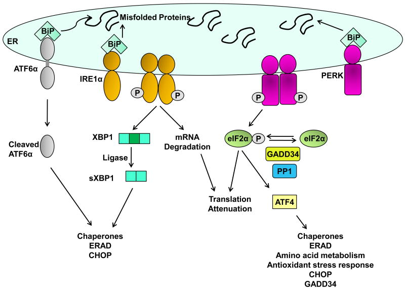
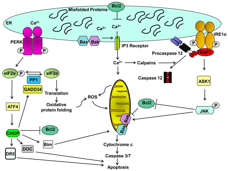
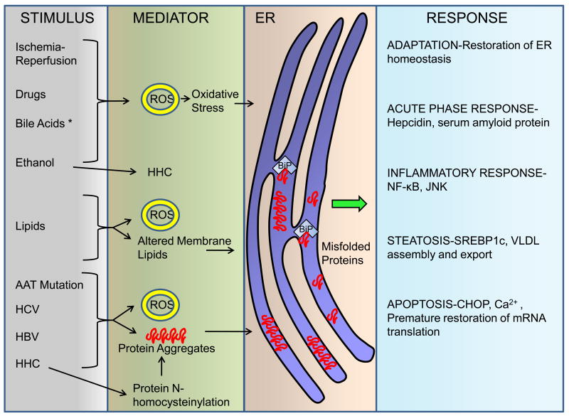
The unfolded protein response (UPR) is activated upon the accumulation of misfolded proteins in the endoplasmic reticulum (ER) that are sensed by the binding immunoglobulin protein (BiP)/glucose-regulated protein 78 (GRP78). The accumulation of unfolded proteins sequesters BiP so it dissociates from three ER-transmembrane transducers leading to their activation. These transducers are inositol requiring (IRE) 1α, PKR-like ER kinase (PERK), and activating transcription factor (ATF) 6α. PERK phosphorylates eukaryotic initiation factor 2 alpha (eIF2α) resulting in global mRNA translation attenuation, and concurrently selectively increases the translation of several mRNAs, including the transcription factor ATF4, and its downstream target CHOP. IRE1α has kinase and endoribonuclease (RNase) activities. IRE1α autophosphorylation activates the RNase activity to splice XBP1 mRNA, to produce the active transcription factor sXBP1. IRE1α activation also recruits and activates the stress kinase JNK. ATF6α transits to the Golgi compartment where it is cleaved by intramembrane proteolysis to generate a soluble active transcription factor. These UPR pathways act in concert to increase ER content, expand the ER protein folding capacity, degrade misfolded proteins, and reduce the load of new proteins entering the ER. All of these are geared toward adaptation to resolve the protein folding defect. Faced with persistent ER stress, adaptation starts to fail and apoptosis occurs, possibly mediated through calcium perturbations, reactive oxygen species, and the proapoptotic transcription factor CHOP. The UPR is activated in several liver diseases; including obesity associated fatty liver disease, viral hepatitis, and alcohol-induced liver injury, all of which are associated with steatosis, raising the possibility that ER stress-dependent alteration in lipid homeostasis is the mechanism that underlies the steatosis. Hepatocyte apoptosis is a pathogenic event in several liver diseases, and may be linked to unresolved ER stress. If this is true, restoration of ER homeostasis prior to ER stress-induced cell death may provide a therapeutic rationale in these diseases. Herein we discuss each branch of the UPR and how they may impact hepatocyte function in different pathologic states.


Conflict of interest statement

Conflict of Interest: The authors declare that they have no conflict of interest with respect to the information presented in this manuscript.

Figures

Figure 1
Figure 1. The unfolded protein response sensors
Three ER membrane sensors activating transcription factor 6 α (ATF6α), inositol requiring (IRE) 1α, and PKR-like ER-localized kinase (PERK) mediate signals from the endoplasmic reticulum (ER) upon activation of the unfolded protein response (UPR). The accumulation of misfolded proteins in the ER lumen sequesters the chaperone BiP away from the lumenal domain of all three ER sensors which leads to their activation. ATF6α is activated by regulated intramembrane proteolysis in the Golgi to release the transcriptionally active 50 kDa cytosolic N-terminal domain. Cleaved ATF6α heterodimerizes with spliced XBP1 (sXBP1) to transcriptionally induce several genes encoding ER chaperones and ER-associated degradation (ERAD) proteins. IRE1α undergoes dimerization and transautophosphorylation which activates its endoribonuclease (RNase) activity. It cleaves X-box binding protein 1 (Xbp1) mRNA, which is then ligated by an uncharacterized ligase to form sXBP1 encoding a potent transcription factor, that also induces expression of ERAD proteins and chaperones. Dimerization and transautophosphorylation of PERK activates its kinase activity, leading to phosphorylation of the alpha subunit of eukaryotic initiation factor 2 (eIF2α). This leads to global translation attenuation. Selective translation of activating transcription factor 4 (ATF4) occurs following phosphorylation of eIF2α. ATF4 induces the expression of several genes including amino acid transporters, chaperones, and C/EBP homologous protein (CHOP). CHOP also induces the expression of GADD34, which associates with protein phosphatase 1 (PP1) to dephosphorylate eIF2α in a negative feedback loop, thus resuming translation.
Figure 2
Figure 2. ER stress-induced apoptosis
Sustained ER stress is associated with cell death. Cells that are dying upon ER stress demonstrate evidence of ongoing UPR, or a lack of resolution of the UPR. Some of the pathways that can lead to ER-stress induced apoptosis are depicted. The proapoptotic proteins Bax and Bak as well as the antiapoptotic protein Bcl-2 are localized on the ER membrane and regulate Ca2+ homeostasis. Calcium release from the ER can activate calpains, which may proteolytically activate caspase 12 to mediate apoptosis. The downstream effectors of caspase 12-induced apoptosis are not known, but presumably promote activation of terminal caspases. Calcium uptake by mitochondria leads to mitochondrial permeabilization and release of cytochrome C. CHOP can induce the expression of proapoptotic BH3-only protein Bim, the cell surface death receptor TRAIL receptor 2, other downstream of chop (DOC) mRNAs, and inhibit Bcl-2 transcription. Oxidative protein folding and mitochondrial dysfunction are associated with the accumulation of reactive oxygen species (ROS) with downstream oxidative cellular damage. JNK is activated by IRE1α via TRAF2. JNK can phosphorylate and activate proapoptotic Bcl-2 family proteins and inactivate antiapoptotic proteins.
Figure 3
Figure 3. ER dysfunction in liver disease
Perturbations in ER homeostasis leading to dysfunction and activation of some of the UPR sensors occur in several liver diseases. Depicted here are the stimuli that can lead to ER dysfunction. The mechanism for some of them includes generation of reactive oxygen species (ROS) leading to oxidative stress, altered membrane lipid composition, hyperhomocysteinemia (HHC) with subsequent protein N-homocysteinylation, formation of protein aggregates, or in some instances are unknown (depicted with an asterisk *). It is likely that additional mediators exist, that are yet to be characterized. In hepatocytes, ER dysfunction leads to many different responses. The UPR is activated to restore ER homeostasis. Steatosis occurs in the liver following acute ER stress, mediated by the lipid-regulatory transcription factors sterol regulatory element binding protein (SREBP)-1c and 2, as well as dysregulation of VLDL assembly and secretion. Inflammatory response cascades are activated in both acute and chronic liver diseases. In alpha-1 antitrypsin (AAT) deficiency the activation of nuclear factor-κB (NF-κB) occurs downstream of perturbations of the ER. Chronic viral hepatitis (hepatitis C virus HCV, hepatitis B virus HBV) is also associated with ER dysfunction. Sustained ER stress leads to apoptosis which may be mediated by C/EBP homologous protein (CHOP), altered Ca2+ homeostasis, and premature resumption of mRNA translation.

References

    1. Asselah T, Bieche I, Mansouri A, Laurendeau I, Cazals-Hatem D, Feldmann G, et al. In vivo hepatic endoplasmic reticulum stress in patients with chronic hepatitis C. J Pathol. 2010;221(3):264–274. - PubMed
    1. Awe K, Lambert C, Prange R. Mammalian BiP controls posttranslational ER translocation of the hepatitis B virus large envelope protein. FEBS Lett. 2008;582(21-22):3179–3184. - PubMed
    1. Back SH, Scheuner D, Han J, Song B, Ribick M, Wang J, et al. Translation attenuation through eIF2alpha phosphorylation prevents oxidative stress and maintains the differentiated state in beta cells. Cell Metab. 2009;10(1):13–26. - PMC - PubMed
    1. Bailly-Maitre B, Belgardt BF, Jordan SD, Coornaert B, von Freyend MJ, Kleinridders A, et al. Hepatic Bax inhibitor-1 inhibits IRE1alpha and protects from obesity-associated insulin resistance and glucose intolerance. J Biol Chem. 2010;285(9):6198–6207. - PMC - PubMed
    1. Bailly-Maitre B, Fondevila C, Kaldas F, Droin N, Luciano F, Ricci JE, et al. Cytoprotective gene bi-1 is required for intrinsic protection from endoplasmic reticulum stress and ischemia-reperfusion injury. Proc Natl Acad Sci U S A. 2006;103(8):2809–2814. - PMC - PubMed

Publication types

MeSH terms