. 2011 Feb 15;350(2):393-404.
doi: 10.1016/j.ydbio.2010.12.007. Epub 2010 Dec 10.

The primitive endoderm lineage of the mouse blastocyst: sequential transcription factor activation and regulation of differentiation by Sox17

Affiliations

The primitive endoderm lineage of the mouse blastocyst: sequential transcription factor activation and regulation of differentiation by Sox17

Jérôme Artus et al. Dev Biol. .

Abstract

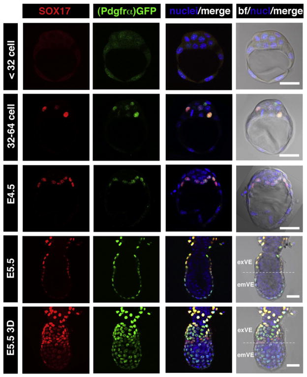
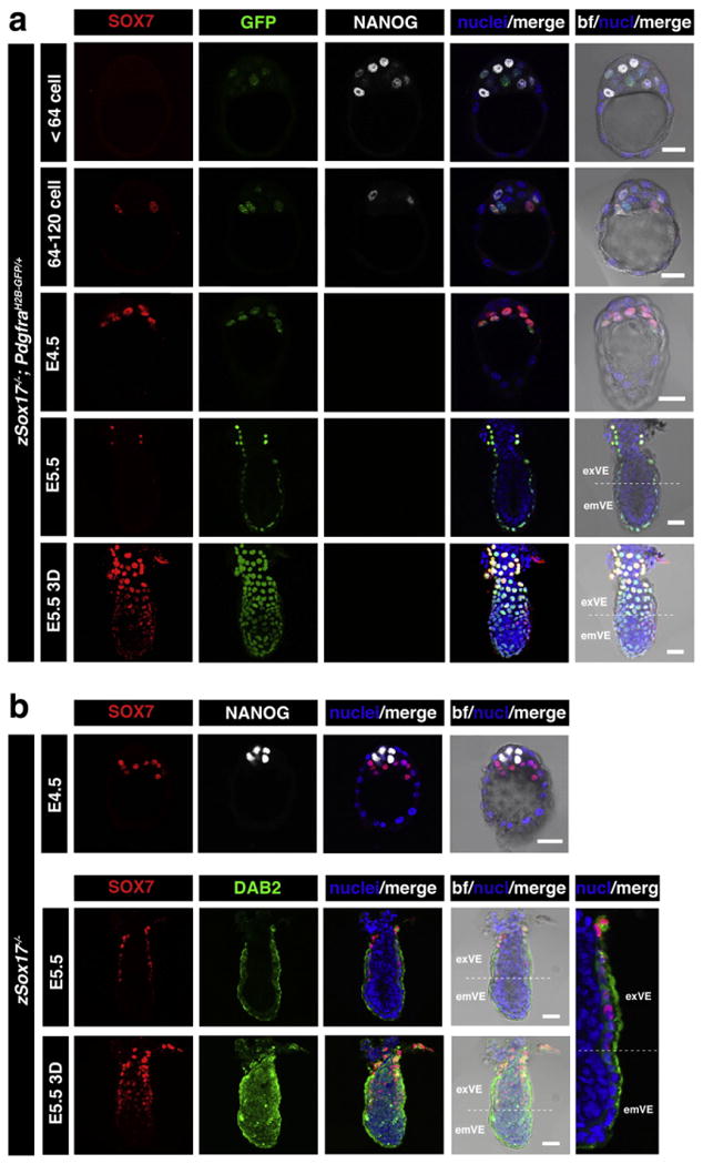
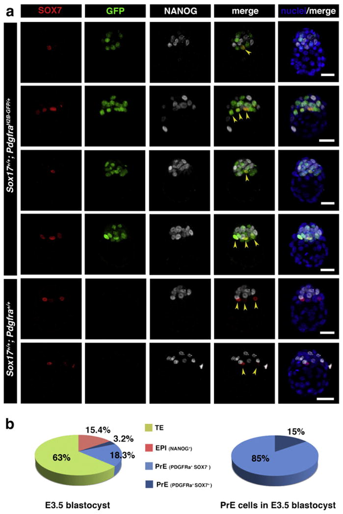
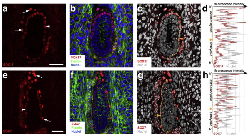
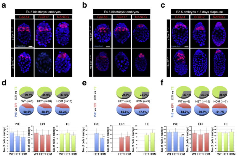
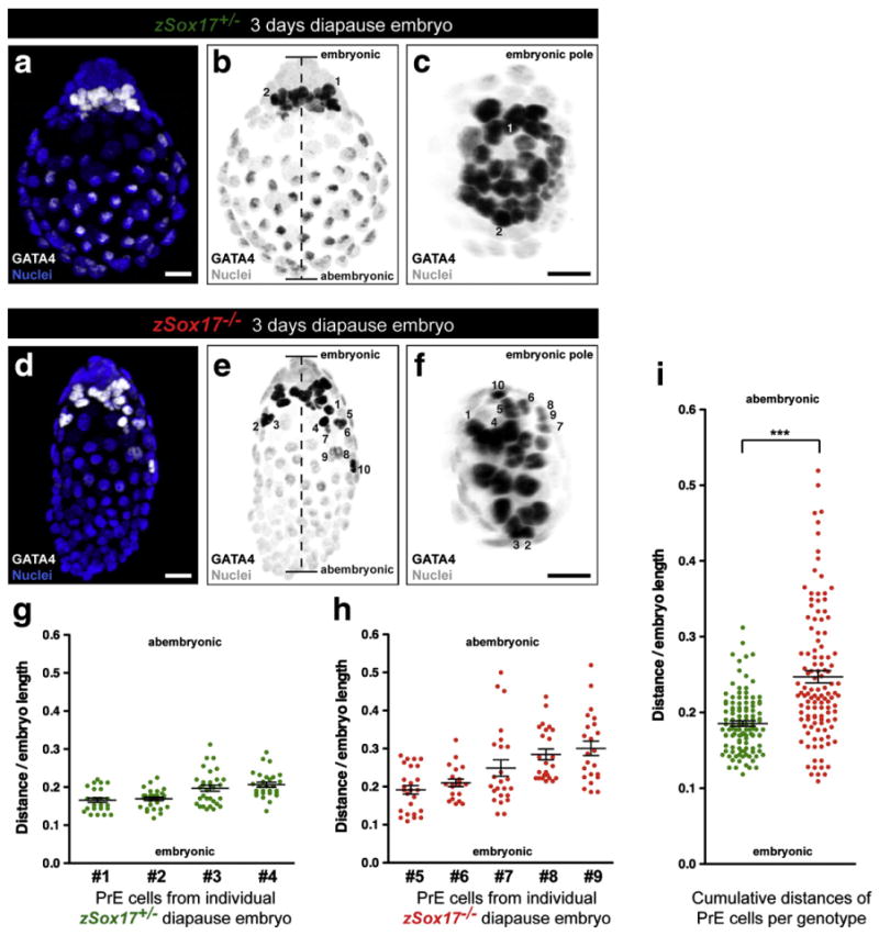
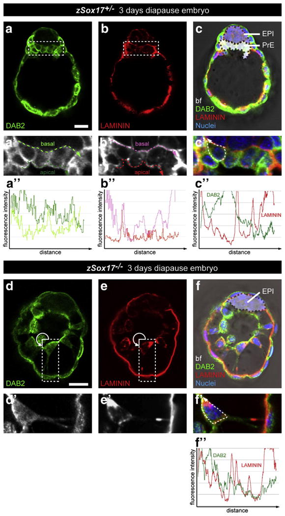
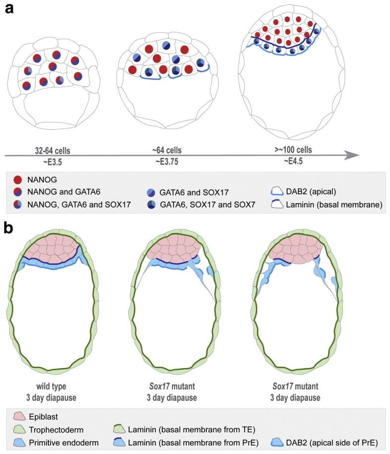
Cells of the primitive endoderm (PrE) and the pluripotent epiblast (EPI), the two lineages specified within the inner cell mass (ICM) of the mouse blastocyst stage embryo, are segregated into adjacent tissue layers by the end of the preimplantation period. The PrE layer which emerges as a polarized epithelium adjacent to the blastocoel, with a basement membrane separating it from the EPI, has two derivatives, the visceral and parietal endoderm. In this study we have investigated the localization of two transcriptional regulators of the SOX family, SOX17 and SOX7, within the PrE and its derivatives. We noted that SOX17 was first detected in a salt-and-pepper distribution within the ICM, subsequently becoming restricted to the nascent PrE epithelium. This dynamic distribution of SOX17 resembled the localization of GATA6 and GATA4, two other PrE lineage-specific transcription factors. By contrast, SOX7 was only detected in PrE cells positioned in contact with the blastocoel, raising the possibility that these cells are molecularly distinct. Our observations support a model of sequential GATA6 > SOX17 > GATA4 > SOX7 transcription factor activation within the PrE lineage, perhaps correlating with the consecutive periods of cell lineage 'naïvete', commitment and sorting. Furthermore our data suggest that co-expression of SOX17 and SOX7 within sorted PrE cells could account for the absence of a detectable phenotype of Sox17 mutant blastocysts. However, analysis of implantation-delayed blastocysts, revealed a role for SOX17 in the maintenance of PrE epithelial integrity, with the absence of SOX17 leading to premature delamination and migration of parietal endoderm.


Figures

Fig. 1
Fig. 1
SOX17 is a marker of the primitive endoderm and its derivatives, the visceral and parietal endoderm of the mouse embryo. SOX17 is first detected in E3.5 embryos at 32–64 cell stage, localizing to GFP expressing cells in PdgfraH2B–GFP/+ embryos, and at E4.5 is found exclusively in the PrE layer. After implantation (E5.5) SOX17 is expressed only in the two PrE derivatives, PE, which exhibits the highest levels of expression, and VE, which exhibits reduced levels of expression towards distal end of the embryo overlying the epiblast. Each horizontal row represents one embryo. All panels depict single optical sections, the lowest panel depicts a 3D-reconstructions of a confocal z-stack. em: embryonic region; ex: extraembryonic region. Pdgfrα-GFP, green; SOX17, red; Hoechst, blue. Scale bar: 20 μm.
Fig. 2
Fig. 2
SOX7 marks PrE cells positioned adjacent to the blastocyst cavity and is expressed in the absence of Sox17. Immunodetection of SOX7 in periimplantation mouse embryos. (a) SOX7 is first detected in E3.5 embryos after 64-cell stage, localizing only to cells in contact with the blastocoel cavity, and in PdgfraH2B–GFP/+ embryos to GFP-positive cells (PrE committed). At E4.5 its expression is restricted to PrE. After implantation (E5.5) SOX7 is detected weakly in exVE. (b) In Sox17−/− embryos, Sox7 expression is not affected at E4.5 and E5.5. Each row represents one embryo. All panels show single optical sections, the lowest panel shows 3D-reconstruction of confocal image. em: embryonic region; ex: extraembryonic region. Pdgfrα-GFP, green (a); DAB2, green (b); SOX7, red; NANOG, white; Hoechst, blue. Scale bar: 20 μm.
Fig. 3
Fig. 3
SOX7 is selectively localized to the PrE cells positioned adjacent to the blastocyst cavity prior to lineage segregation. (a) Immunodetection of SOX7 in preimplantation mouse embryos at E3.5 (65–80 cells). SOX7 is first detected in E3.5 embryos after 64-cell stage, localizing only to cells in contact with the blastocoel cavity (arrowheads). In PdgfraH2B–GFP/+ embryos it is co-localized with GFP in cells adjacent to the cavity. Each row represents one embryo, all panels show 3D-reconstruction of confocal images. Pdgfrα-GFP, green; SOX7, red; NANOG, white; Hoechst, blue. Scale bar: 20 μm. (b) Distribution of TE (green), EPI (red) and PrE (SOX7-negative — light blue, and SOX7-positive — dark blue) cells in embryos at E3.5.
Fig. 4
Fig. 4
SOX7 and SOX17 are detected in PrE derivatives at E5.5 postimplantation stage. Immunodetection of SOX17 (a–d) and SOX7 (e–h) on cryosections of decidua containing E5.5 wild-type embryos. SOX17 and SOX7 were detected both in PE (arrows) and VE (arrowheads). (d, h) Measurement of fluorescence intensity of SOX17 (d) and SOX7 (h) along the proximal–distal axis. Orange dashed lines indicate where the measurements were done and orange arrowheads indicate the boundary between embryonic and abembryonic regions. SOX7, SOX17, red; nuclei, blue and grey; F-actin, green. Scale bar: 50 μm.
Fig. 5
Fig. 5
Mouse blastocysts lacking Sox17 correctly specify and segregate embryonic and extraembryonic lineages. Zygotically zSox17 +/− (upper panels) and zSox17−/− embryos (lower panels) at E4.5 (a) and 3 days after tamoxifen injection (c). Maternally and zygotically ablated mzSox17 +/− (upper panels) and mzSox17−/− embryos (lower panels) at E4.5 (b), Blue, nuclei; red, GATA4, GATA6, SOX17 and NANOG. Scale bar: 20 μm. (d–f) Distribution of PrE, EPI and TE cells in Sox17+/+, Sox17+/−, Sox17−/− embryos at E4.5 (d), 3 days after tamoxifen injection (f) and in maternal/zygotic mutant embryos at E4.5 and (e). Blue, PrE; red, EPI; green, TE; grey, ICM. Statistical T-tests are indicated when significant (*:p<0.02; **:p<0.003). Error bars indicate s.d.
Fig. 6
Fig. 6
Premature migration of PrE cells along mural trophectoderm in implantation delayed Sox17 mutant blastocysts. (a, d) Immunodetection of GATA4 (white) in 3 day implantation-delayed Sox17+/− (a) and Sox17−/− embryos (d). Nuclei were counterstained with Hoechst (blue). (b, e) Greyscale conversion of (a) and (d) respectively. Dashed line defines the length between the embryonic and abembryonic poles. (c, f) View from the embryonic pole of (b) and (e) respectively. GATA4-positive nuclei from PrE cells (dark, number) can be tracked and located according to these two views. (a–f) Scale bars: 20 μm. (g) Average and relative distances to the proximal embryonic region of the PrE cells in 4 individual Sox17+/− (green) embryos. (h) Average and relative distances to the proximal embryonic region of the PrE cells in 5 individual Sox17−/− (red) embryos. (i) Combined average and relative distances of PrE cells in all analyzed Sox17+/− (green) and Sox17−/− (red) embryos. (***: significant statistical t-test with a p<0.0001). (g–i) Error bars indicate s.e.m.
Fig. 7
Fig. 7
Disrupted organization of primitive endoderm correlates with basement membrane deposition in the absence of Sox17. Immunodetection of DAB2 and Laminin in Sox17+/− (a–c, a′–c′, a″–c″) and Sox17−/− (d–f, d′–f′, f″) 3 day diapause embryos. (a′–f′) magnified views of (a–f) respectively. (a″,b″) Measurements of the fluorescence intensity of DAB2 (a″) and Laminin (b″) staining along the apical (green and red dashed lines) and basal (light green and pink dashed lines) of the PrE layer. (c″,f″) Measurements of the fluorescence intensity of Laminin (red) and DAB2 (green) throughout the cell membrane (dashed line). All panels show single optical section. DAB2, green; Laminin, red; nuclei, blue; bf, bright field. Scale bar: 20 μm.
Fig. 8
Fig. 8
Schematic representations of sequential activation of transcription factors in the PrE cell lineage and the phenotype resulting from inactivation of Sox17 in implantation-delayed blastocysts. (a) At E3.5, in embryos of 32–64 cells, GATA6 is expressed in all cells of the ICM, and SOX17 can be detected in some cells of the ICM. At around the 64-cell stage, coincident with the emergence of salt-and-pepper distribution of EPI/PrE cells, GATA6 becomes downregulated in presumptive EPI cells, and SOX17 can be detected in most of these presumptive PrE cells, such that GATA6 and SOX17 co-localize. SOX7 is localized to GATA6/SOX17-positive cells that are positioned on the cavity roof. These are the cells that will, or are just beginning to, accumulate DAB2 apically (Gerbe et al., 2008). At the time of implantation (E4.5), the PrE is apparent as a morphologically-distinct epithelial cell layer in contact to the cavity with cells being GATA6/SOX17/SOX7-positive. (b) Inactivation of Sox17 in implantation-delayed blastocyst stage embryos results in disruption of the organization of the PrE epithelium and premature parietal endoderm differentiation. Premature disattachment and migration of nascent parietal endoderm cells over the TE layer are observed. Wild-type embryo is depicted on the left, middle panel represents a Sox17 mutant exhibiting a mild defect, whereas the right panel depicts a mutant exhibiting a more severe defect.

References

    1. Adamson ED, Ayers SE. The localization and synthesis of some collagen types in developing mouse embryos. Cell. 1979;16:953–965. - PubMed
    1. Alexander J, Stainier DY. A molecular pathway leading to endoderm formation in zebrafish. Curr Biol. 1999;9:1147–1157. - PubMed
    1. Artus J, Panthier JJ, Hadjantonakis AK. A role for PDGF signaling in expansion of the extra-embryonic endoderm lineage of the mouse blastocyst. Development. 2010;137:3361–3372. - PMC - PubMed
    1. Bowles J, Schepers G, Koopman P. Phylogeny of the SOX family of developmental transcription factors based on sequence and structural indicators. Dev Biol. 2000;227:239–255. - PubMed
    1. Chazaud C, Yamanaka Y, Pawson T, Rossant J. Early lineage segregation between epiblast and primitive endoderm in mouse blastocysts through the Grb2–MAPK pathway. Dev Cell. 2006;10:615–624. - PubMed

Publication types