. 2011 Feb 15;350(2):370-81.
doi: 10.1016/j.ydbio.2010.12.005. Epub 2010 Dec 10.

C. elegans Dicer interacts with the P-granule component GLH-1 and both regulate germline RNPs

Affiliations

C. elegans Dicer interacts with the P-granule component GLH-1 and both regulate germline RNPs

Erica L Beshore et al. Dev Biol. .

Abstract

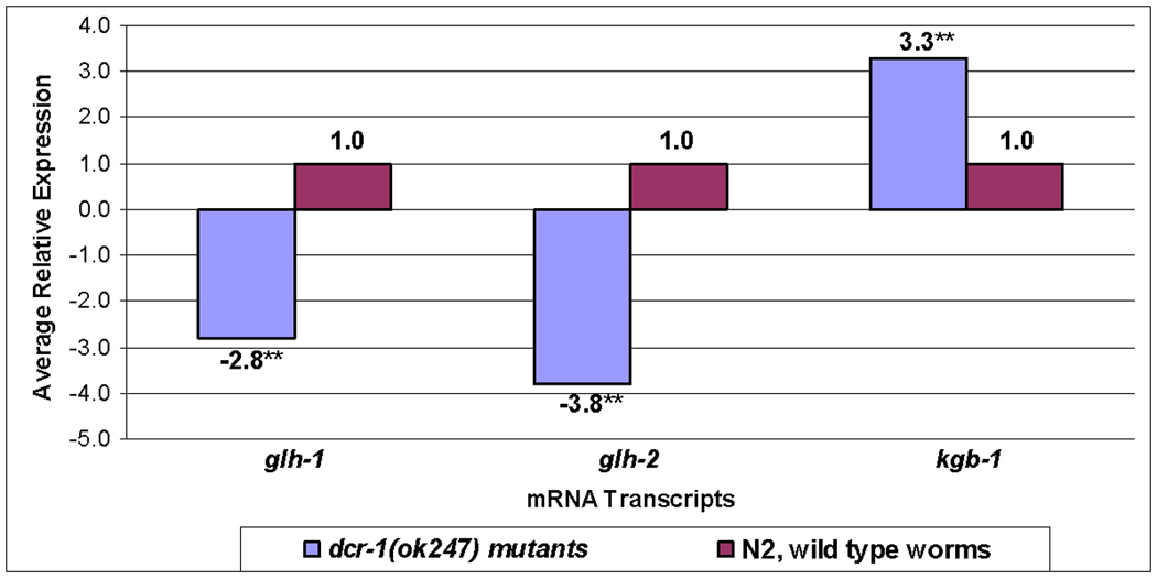
P granules, ribonucleoprotein (RNP) complexes specific to the cytoplasmic side of the nuclear pores of Caenorhabditis elegans germ cells, are implicated in post-transcriptional control of maternally-transcribed mRNAs. Here we show a relationship in C. elegans of Dicer, the riboendonuclease processing enzyme of the RNA interference and microRNA pathways, with GLH-1, a germline-specific RNA helicase and a constitutive component of P granules. Based on results from GST-pull-downs and immunoprecipitations, GLH-1 binds DCR-1 and this binding does not require RNA. Both GLH-1 protein and glh-1 mRNA levels are reduced in the dcr-1(ok247) null mutant background; conversely, a reduction of DCR-1 protein is observed in the glh-1(gk100) deletion strain. Thus, in the C. elegans germline, DCR-1 and GLH-1 are interdependent. In addition, evidence indicates that DCR-1 protein levels, like those of GLH-1, are likely regulated by the Jun N-terminal kinase (JNK), KGB-1. In adult germ cells, DCR-1 is found in uniformly-distributed, small puncta both throughout the cytoplasm and the nucleus, on the inner side of nuclear pores, and associated with P granules. In arrested oocytes, GLH-1 and DCR-1 re-localize to cytoplasmic and cortically-distributed RNP granules and are necessary to recruit other components to these complexes. We predict that the GLH-1/DCR-1 complex may function in the transport, deposition, or regulation of maternally-transcribed mRNAs and their associated miRNAs.


Figures

Figure 1
Figure 1. GLH-1 interacts with DCR-1 and PGL-1
A. A cartoon summarizing the interactions seen in Figs. 1B–C. B. Pull-downs out of wild type worm lysates with full length GLH-1 (lane 1), GST alone (lane 2), N-GLH-1 (lane 3), N*-GLH-1 (lane 4), C-GLH-1 (lane 5), full-length GLH-1, after adding RNase A (12 µg/µL for 20 min at room temperature) to the lysate prior to pull-down (lane 6), full-length GLH-1 without RNase A (lane 7), and 10% input lysate (lane 8). These pull-downs were tested with α-DCR-1 antibodies. C. Pull-downs out of wild type worm lysates with full length GLH-1 (lane 1), N-GLH-1 (lane 2), GST alone (lane 3), C-GLH-1 (lane 4), full-length GLH-1 after adding RNase A, as in Fig. 1B, to the lysate prior to pull-down (lane 5), full-length GLH-1 without RNase A, but treated as in lane 5, (lane 6), and 10% input lysate (lane 7). All GLH-1 proteins were GST-tagged except C-GLH-1, which was HIS-tagged. All lanes were tested by western blot with α-PGL-1 antibodies. The PGL-1 protein in lane 1 is no smaller than in other lanes; the gel edges ran slightly further. Western blots with α-GST and α-HIS antibodies show proteins were present in all lanes, Fig. S1.
Figure 2
Figure 2. DCR-1 localizes in close proximity to nuclear pore complexes
A. A schematic of the C. elegans gonad from an adult hermaphrodite containing the adult germline, adapted from Minasaki et al. (2009). In the distal gonad, the somatic distal tip cell directs mitotic germ cell proliferation; germ cells then transition into meiosis and enter pachytene. The distal region is a syncytium, while in the proximal gonad oocytes cellularize and mature before passing through the spermatheca where they are fertilized. B. DCR-1 (green) is localized throughout the adult gonad in a wild type hermaphrodite, both throughout the cytoplasm and in the P granules surrounding the germ cell nuclei. This image of a splayed gonad includes some distal and proximal regions surrounding the gonad bend. The same gonad is seen in the middle panel, showing the localization of GLH-1 (red). DCR-1 and GLH-1 appear to co-localize in the merged image (yellow). These images were taken with a Zeiss Axioplan microscope. Size markers=25 µm. C. Confocal micrographs of the pachytene region of a wild type hermaphrodite using mAb414 antibody (green), which recognizes the FG repeats of several nucleoporins (Davis and Blobel, 1986) and α-DCR-1 antibody (red). D. Immunocytochemistry in the pachytene region of a wild type hermaphrodite showing α-GLH-1 (red), α-DCR-1 (green) and mAb414 (blue) antibodies. E. Immunocytochemistry in a wild type oocyte showing α-GLH-1 (red), α-DCR-1 (green), and mAb414 (blue) antibodies. Arrows denote the region enlarged in each inset. Size markers=5 µm. To evaluate the extent of co-localization between pairs of proteins in co-staining experiments, Pearson’s Correlation was determined using Nikon NIS Elements software. Pearson’s Correlation is a commonly accepted means for describing the extent of overlap between image pairs and takes into account only the similarity of shapes between images but not the image intensity (Adler et al., 2010).
Figure 3
Figure 3. GLH-1 and DCR-1 are interdependent
A. Western blot analysis of wild type versus dcr-1(ok247) protein lysates with α-GLH-1 antibody; tubulin is the loading control. B. Western blot analysis of wild type versus glh-1(gk100) protein lysates with α-DCR-1 antibody; actin is the loading control. The western blot in 3A used 50 worms per lane, while those in 3B used 500 worms per lane to compensate for the low levels (~10%) of truncated GLH-1 protein in the glh-1(gk100) strain. The use of different loading controls between the western blots in A and B was unintended, reflecting the availability of these control antibodies at the time. C. GLH-1 localizes to P granules in wild type (left); GLH-1 is not detected in a dcr-1(ok247) gonad (right) using α-GLH-1 (red), and DAPI-stained nuclei (blue). D. A wild type and a dcr-1(ok247) gonad reacted with α-PGL-1 antibody (green), and DAPI-stained nuclei (blue). Arrows indicate the distal gonad. The arrowhead points to two endomitotic oocytes (Emo). E. A glh-1(gk100) gonad and intestine reacted with α-DCR-1 (green) and stained with DAPI (blue). The arrowhead points to a small section of the C. elegans intestine. The arrow indicates the gonad distal tip. F. A wild type gonad with α-DCR-1 (green) and DAPI (blue). All worms were grown at 20°C. Size markers=20 µm, except 3F, which =10 µm.
Figure 4
Figure 4. glh-1 transcript levels are reduced about 3 fold in dcr-1(ok247) when compared to wild type worms
Transcript levels of glh-1, glh-2, and kgb-1 were assayed in triplicate by qRT-PCR in dcr-1(247) as compared to wild type worms (**p value<0.001). The statistical significance of differences in relative mRNA levels was determined using the Relative Expression Software Tool (REST) 2009 (www.gene-quantification.de/rest.html). The primers used are shown in Table S1 and the numerical results are given in Table S2.
Figure 5
Figure 5. DCR-1 is elevated when JNK pathways are inhibited and DCR-1 levels accumulate in kgb-1 worms
A. Wild type worms were treated with 50 µM of the JNK inhibitor, SP600125, which was added to liquid culture media. Fifty worms treated with inhibitor for 0, 1, 2, 4 or 6 hours were collected after all had grown for 6 hrs and then analyzed by western blot for DCR-1. GLH-4 was a loading control since GLH-4 levels do not change in kgb-1(um3) worms (Orsborn et al., 2007). This image was recombined, removing the 2 and 4 hour time points, which also showed increasing DCR-1 protein levels; however, the GLH-4 control did not transfer well in that region of the membrane. B. Levels of DCR-1 were compared by western blot analysis in age-matched wild type and kgb-1(um3) adults grown at 26°C. Actin was the loading control. C. The pachytene region of a kgb-1(um3) mutant, grown at 20°C, reacted with antibodies against DCR-1 (green), and GLH-1 (red), merged. Similar strongly-reactive, large P granules were previously reported for GLH-1 in kgb-1(um3) worms (Smith et al., 2002). D. The pachytene region of a wild type gonad, with α-DCR-1 (green) and α-GLH-1 (red). E–F. DAPI-stained (blue) gonads in panels C–D, respectively. G. A section of intestine from a kgb-1(um3) worm with α-DCR-1 (green). H. A section of intestine from a wild type worm, with α-DCR-1 (green). I. Merged image of a P3-stage (8-cell) kgb-1(um3) embryo with antibodies again DCR-1 (green), GLH-1 (red), and DAPI-stained nuclei (blue). J. Merged image of a P4-stage (16-cell) wild type embryo reacted with antibodies against DCR-1 (green), GLH-1 (red), and DAPI-stained nuclei (blue). The P3 and P4 germ cell progenitors are marked (arrowheads). Each set of images comparing kgb-1(um3) and wild type worms (C/D; G/H or I/J) were taken with the same settings and exposures. Worms were grown at 20°C unless otherwise noted. Size markers=10 µm.
Figure 6
Figure 6. DCR-1 and GLH-1 localize to RNP granules
A. Confocal single plane images of the five most-proximal fog-2(q71) arrested oocytes, reacted with antibodies against GLH-1 (green), DCR-1 (red), and merged. B. The gonad core of a fog-2(q71) unmated female, with antibodies against nuclear pores (left, blue), DCR-1 (middle, green), and GLH-1 (right, red). Combined localizations are seen with merged images (bottom). Size markers=5 µm.
Figure 7
Figure 7. Reduction of GLH-1 or DCR-1 disrupts assembly of the cytoplasmic RNP granules
MEX-3 (A–E) and PGL-1 (F–H) were visualized with their respective antibodies. Unmated fog-2(q71) females were the control for normal RNP granule assembly (A, F). MEX-3 and PGL-1 failed to localize to large granules in glh-1(gk100) worms depleted of sperm (B, G); however, GLH-1 was not essential for MEX-3 to localize to RNP granules after heat shock (C). An intermediate effect was seen for PGL-1 (H). DCR-1 appeared to regulate the localization of MEX-3 to RNP granules after heat shock and when sperm are depleted (D–E). Size markers=5 µm.
Figure 8
Figure 8. Model of the GLH-1/DCR-1 relationship in P granules and RNP granules
In wild type worms, GLH-1 and DCR-1 localize to P granules to regulate the fate of maternal mRNAs and their associated miRNAs. Both proteins are subject to degradation by the proteasome, which is indicated by a PacMan© image. Under conditions of arrested ovulation, GLH-1 and DCR-1 re-localize to large RNP granules to store maternal mRNAs and recruit MEX-3, PGL-1, and perhaps CGH-1. Nuclear pores are indicated with blue ovals; miRNAs (short red lines) are located on the 3’UTRs of their target mRNAs (long black lines).

References

    1. Adler J, Parmryd I. Quantifying co-localization by correlation: the Pearson correlation coefficient is superior to the Mander's overlap coefficient. Cytometry Part A. 2010;77A:733–742. - PubMed
    1. Anderson P, Kedersha N. RNA granules. J. Cell Biol. 2006;172(6):803–808. Mini-Review. - PMC - PubMed
    1. Amiri A, Keiper BD, Kawasaki I, et al. An isoform of eIF4E is a component of germ granules and is required for spermatogenesis in C. elegans. Development. 2001;128:3899–3912. - PMC - PubMed
    1. Balagopal V, Parker R. Polysomes, P bodies and stress granules: states and fates of eukaryotic mRNAs. Curr. Opin. Cell Biol. 2009;21(3):403–408. - PMC - PubMed
    1. Barbee SA, Lubin AL, Evans TC. A novel function for the Sm proteins in germ granule localization during C. elegans embryogenesis. Curr. Biol. 2002;12:1502–1506. - PubMed

Publication types

MeSH terms