. 2011 Feb 25;406(3):371-86.
doi: 10.1016/j.jmb.2010.11.027. Epub 2010 Dec 10.

Structure of the HIV-1 full-length capsid protein in a conformationally trapped unassembled state induced by small-molecule binding

Affiliations

Structure of the HIV-1 full-length capsid protein in a conformationally trapped unassembled state induced by small-molecule binding

Shoucheng Du et al. J Mol Biol. .

Abstract

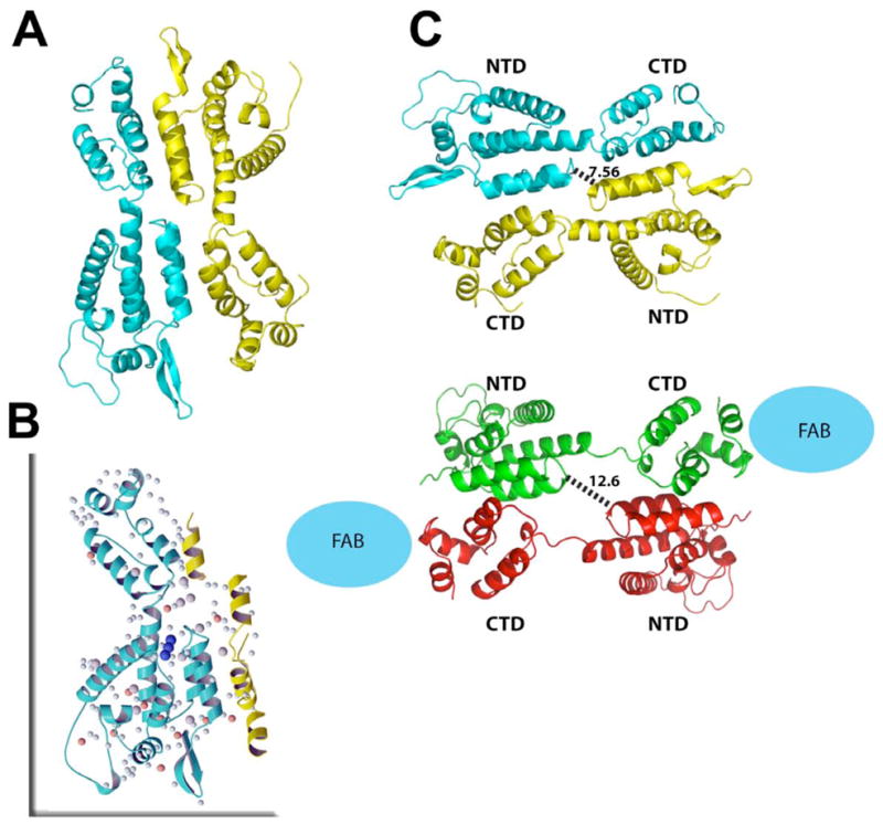
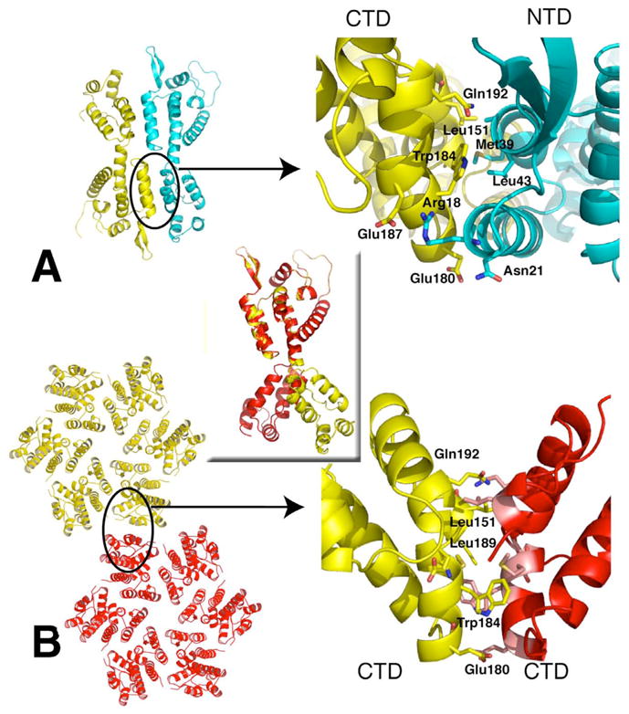
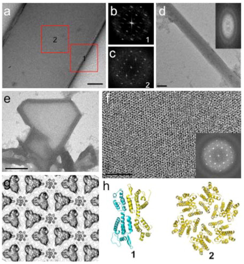
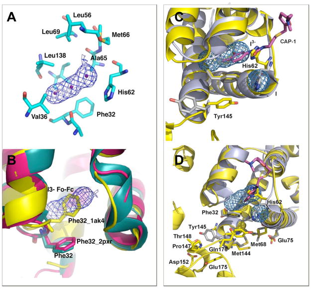
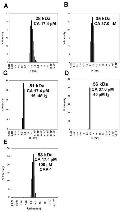
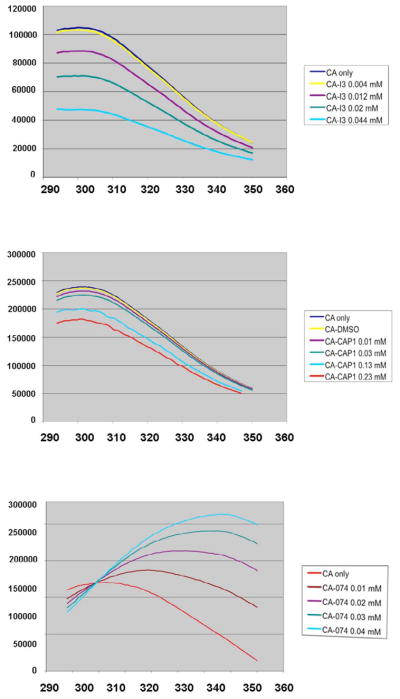
The capsid (CA) protein plays crucial roles in HIV infection and replication, essential to viral maturation. The absence of high-resolution structural data on unassembled CA hinders the development of antivirals effective in inhibiting assembly. Unlike enzymes that have targetable, functional substrate-binding sites, the CA does not have a known site that affects catalytic or other innate activity, which can be more readily targeted in drug development efforts. We report the crystal structure of the HIV-1 CA, revealing the domain organization in the context of the wild-type full-length (FL) unassembled CA. The FL CA adopts an antiparallel dimer configuration, exhibiting a domain organization sterically incompatible with capsid assembly. A small compound, generated in situ during crystallization, is bound tightly at a hinge site ("H site"), indicating that binding at this interdomain region stabilizes the ADP conformation. Electron microscopy studies on nascent crystals reveal both dimeric and hexameric lattices coexisting within a single condition, in agreement with the interconvertibility of oligomeric forms and supporting the feasibility of promoting assembly-incompetent dimeric states. Solution characterization in the presence of the H-site ligand shows predominantly unassembled dimeric CA, even under conditions that promote assembly. Our structure elucidation of the HIV-1 FL CA and characterization of a potential allosteric binding site provides three-dimensional views of an assembly-defective conformation, a state targeted in, and thus directly relevant to, inhibitor development. Based on our findings, we propose an unprecedented means of preventing CA assembly, by "conformationally trapping" CA in assembly-incompetent conformational states induced by H-site binding.


Figures

Figure 1
Figure 1
Crystal asymmetric unit (A) with one monomer shown in yellow, one in cyan. Extensive solvent and ion network (B) comprised of irons (orange spheres), iodides (larger gray spheres) and waters (small light cyan spheres) mediate interactions between domains within the FL CA (in blue ribbon) and between protein subunits (neighboring subunit partially shown in yellow ribbon). Binding of triiodide (dark blue spheres) close to the hinge likes stabilizes the conformation. Comparison between the native full-length CA structure to the RH24 complexed to a Fab fragment (C) with native FL CA dimer in cyan and yellow and the RH24 dimer in green and red ribbons. The antiparallel dimer in both structures exhibit similar molecular organization but in RH24, the two halves of the dimer are stretched further apart because of unraveling of 310 helical bridge between the NTD to CTD, possibly due to Fab (blue sphere) binding.
Figure 2
Figure 2
Relationships between CA domains in the FL CA compared to assembled CA. Crystal asymmetric unit with one monomer shown in yellow the other in cyan (A). Close up view of the CTD-NTD interface with key residues labeled (top right). CryoEM reconstructed pseudo-atomic model from (3DIK) showing two hexamers (B). Proposed hexamer-hexamer interface based on modeling into EM maps isolated CTD dimer structures. Many of the key residues forming the CTD-CTD dimer interface in the EM model are also form NTD-CTD interactions in the FL CA structures. Rotation of 180 degrees is required to superimpose the CTD of a monomer subunit of the FL CA (yellow) onto the hexamer monomer (red; central inset).
Figure 3
Figure 3
EM Analysis on Crystal Nuclei Showing initial 3D Crystals Coexisting with Multiple-layered Tubes at their Edges. A projection image (a) of a small negatively stained 3D crystal after 12 hours crystallization. Fourier transforms from the crystal sheet (b, region 1) and from tubular region (c, region 2); respective models of presumed oligomers representing the diffraction images shown at bottom (h). A projection image (d) and its Fourier transform (insert) of a negatively stained multiplelayered tubular crystal. A low magnification image of a 2D crystal (e) edged with tubes and an enlarged view of the 2D crystalline sheet (f). The Fourier transforms of the 2D crystalline sheet (insert of f) and corresponding projection map (g) indicate packing of hexagonal lattice. Scale bars, 100nm in a and d; 50nm in f, 1um in e.
Figure 4
Figure 4
The Triiodide (I3) and inhibitor, CAP-1, Binding Sites are Located at the Inter Domain Hydrophobic Pocket Formed Between the NTD-CTD. (A) A monomer of FL CA is shown in cyan, with dark blue residues labeled to show the hydrophobic side chains (except His64) surrounding the I3, the difference density map at the site is shown (Fo-Fc, contoured at 6σ). (B) The position of Phe32 in the I3 bound crystal is shown in cyan, in CAP-1 NTD in magenta (2PXR) and the position of Phe32 in unliganded NTD structures is shown in yellow (1AK45). (C) A striking rotation of Tyr145 is found in the CAP-1 complex, where the aromatic ring is swung out of the pocket. (D) In the I3 complexed structure, Tyr-145 remains associated with the binding pocket, forming interactions to Gln176 and Met144, with each side-chains forming additional networks of H-bond interactions that bridge NTD and CTD, stabilizing domain orientation.
Figure 5
Figure 5
Light Scattering Analysis of CA in Solution in Absence and Presence of I3 & CAP-1. To characterize the solution oligomeric state of CA in the absence and presence of I3, a series of dynamic light scattering analysis were conducted. All measurements were done in 50 mM sodium phosphate buffer at pH 7.0. (A) CA at 17.4 μM concentration is monomeric; (B) CA at 37 μM is predominantly monomeric at low salt; (C) CA at 17 μM concentration with 16 μM I3; (D) CA at 37 μM and 40 μM I3- resulted in the enhanced formation of the dimeric state, even at 17 μM concentration; (E) CA at 17 μM concentration with 100 μM CAP-1.
Figure 6
Figure 6
Fluorescence Analysis of FL CA in Presence of Inhibitors The effects of I3 binding to assembly were characterized by fluorescence spectroscopy to determine if intermolecular associations were affected by binding and compared to two capsid assembly inhibitors, CAP-1 and a Pfizer compound PF-03450074, referred to as “074”. These results are consistent with the reported mechanism of inhibition of 074, which accelerates dissociation of CA to monomers, which rapidly aggregates to protect the hydrophobic residues. The similarity in the fluorescence emission profile of CA in the presence of I3 and CAP-1 are also consistent with the structural results, which identified their binding sites to both be at the interdomain hinge region, supporting the idea that their binding promotes a NTD-CTD domain orientation in the FL CA that stabilizes a dimeric conformation that is not competent for assembly, such as the APD configuration.

References

    1. Ganser-Pornillos BK, Yeager M, Sundquist WI. The structural biology of HIV assembly. Curr Opin Struct Biol. 2008;18:203–17. - PMC - PubMed
    1. Chang YF, Wang SM, Huang KJ, Wang CT. Mutations in capsid major homology region affect assembly and membrane affinity of HIV-1 Gag. J Mol Biol. 2007;370:585–97. - PubMed
    1. Chu HH, Chang YF, Wang CT. Mutations in the alpha-helix directly C-terminal to the major homology region of human immunodeficiency virus type 1 capsid protein disrupt Gag multimerization and markedly impair virus particle production. J Biomed Sci. 2006;13:645–56. - PubMed
    1. Cardone G, Purdy JG, Cheng N, Craven RC, Steven AC. Visualization of a missing link in retrovirus capsid assembly. Nature. 2009;457:694–8. - PMC - PubMed
    1. Ganser-Pornillos BK, Cheng A, Yeager M. Structure of full-length HIV-1 CA: a model for the mature capsid lattice. Cell. 2007;131:70–9. - PubMed

Publication types

Substances

LinkOut - more resources