. 2011 May;275(1-2):66-80.
doi: 10.1016/j.heares.2010.12.002. Epub 2010 Dec 10.

Conditional deletion of Atoh1 using Pax2-Cre results in viable mice without differentiated cochlear hair cells that have lost most of the organ of Corti

Affiliations

Conditional deletion of Atoh1 using Pax2-Cre results in viable mice without differentiated cochlear hair cells that have lost most of the organ of Corti

Ning Pan et al. Hear Res. 2011 May.

Abstract

Atonal homolog1 (Atoh1, formerly Math1) is a crucial bHLH transcription factor for inner ear hair cell differentiation. Its absence in embryos results in complete absence of mature hair cells at birth and its misexpression can generate extra hair cells. Thus Atoh1 may be both necessary and sufficient for hair cell differentiation in the ear. Atoh1 null mice die at birth and have some undifferentiated cells in sensory epithelia carrying Atoh1 markers. The fate of these undifferentiated cells in neonates is unknown due to lethality. We use Tg(Pax2-Cre) to delete floxed Atoh1 in the inner ear. This generates viable conditional knockout (CKO) mice for studying the postnatal development of the inner ear without differentiated hair cells. Using in situ hybridization we find that Tg(Pax2-Cre) recombines the floxed Atoh1 prior to detectable Atoh1 expression. Only the posterior canal crista has Atoh1 expressing hair cells due to incomplete recombination. Most of the organ of Corti cells are lost in CKO mice via late embryonic cell death. Marker genes indicate that the organ of Corti is reduced to two rows of cells wedged between flanking markers of the organ of Corti (Fgf10 and Bmp4). These two rows of cells (instead of five rows of supporting cells) are positive for Prox1 in neonates. By postnatal day 14 (P14), the remaining cells of the organ of Corti are transformed into a flat epithelium with no distinction of any specific cell type. However, some of the remaining organ of Corti cells express Myo7a at late postnatal stages and are innervated by remaining afferent fibers. Initial growth of afferents and efferents in embryos shows no difference between control mice and Tg(Pax2-Cre)::Atoh1 CKO mice. Most afferents and efferents are lost in the CKO mutant before birth, except for the apex and few fibers in the base. Afferents focus their projections on patches that express the prosensory specifying gene, Sox2. This pattern of innervation by sensory neurons is maintained at least until P14, but fibers target the few Myo7a positive cells found in later stages.


Figures

Figure 1
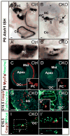
Figure 1. Conditional deletion of Atoh1 is early, almost complete, and affects other bHLH genes but not early neuronal development
(A, B) The Pax2-Cre mediated recombination of floxed Atoh1 leads to reduction and loss of Atoh1 expression as shown by in situ hybridization. In E13.5 control mice, Atoh1 is expressed in all five vestibular endorgans with limited expression in the cochlea (A). In Atoh1 CKO mice, only some weak expression persists in the posterior crista and the anterior crista at this stage (B). (CH)Absence of Atoh1 leads to expression changes in other bHLH genes. At E13.5 Neurog1 shows a wider expression in the utricle and saccule of Atoh1 CKO mice (D) compared to their control littermates (C). At E14.5, while Neurog1 is drastically downregulated in the control mice (G), it continues to be expressed in the CKO mice (H). An expanded expression is also seen for Neurod1 in the mutant ear at E14.5 (E, F). (IJ′) Nhlh2, a neuronal marker shows near normal expression in developing ganglia in CKO mutants. However, the sizes of vestibular ganglia are obviously different: the superior vestibular ganglion is reduced but the inferior vestibular ganglion is enlarged in CKO mutants compared to control littermates (compare the bars in I′ and J′). (K–L′) Lipophilic dye tracing shows that both the afferent and the efferent innervation in the Atoh1 CKO mice (L, L′) is indistinguishable from the control littermate (K, K′). AC, anterior canal crista; Co, cochlea; HC, horizontal canal crista; IGSB, intraganglionic spiral bundle; IVG, inferior vestibular ganglion; PC, posterior canal crista; S, saccule; SG, spiral ganglion; SVG, superior vestibular ganglion; U, utricle. Bar indicates 100 μm.
Figure 2
Figure 2. Loss of Atoh1 causes failure of hair cell differentiation and cell deaths of organ of Corti precursors and neurons
(AB′) Atoh1 in situ hybridization at P0 confirms the deletion of Atoh1 in almost all the hair cells of the mutant ear. The control ear has strong Atoh1 signals in all sensory epithelia (A, A′). In the CKO mutant, there is only some residual expression in the posterior canal crista (arrows in B and B′). The black patches in anterior canal crista, posterior canal crista, utricle and saccule are the pigments but not Atoh1 signal, which shows dark purple color. (CF) Myo7a immunochemistry shows complete loss of hair cells in the mutant cochlea. Consistent with the remaining Atoh1 expression, a few Myo7a positive hair cells are present in the posterior canal crista (F). (GI′) Absence of Atoh1 causes cell deaths of neurons and cells in the organ of Corti. In E18.5 mutant, activated Caspase3 positive cells are seen in spiral ganglion (G) and the region topographically equivalent to the organ of Corti (H). Degeneration of these cells is further evidenced by Hoechst staining of the pyknotic nuclei (H′). In E16.5 mutant, we find a streak of cells in the organ of Corti that are labeled by PSVue, which stains phosphatidylserine exposed on the cell membranes after the onset of apoptotic cell deaths (I). In I′, z-stack confocal image is optically cross sectioned through the organ of Corti (indicated by the dotted lines) and the lateral views shown on the bottom and the side reveal that PSVue positive cells are located in two cell layers, one near the lumen and one near the basilar membrane suggesting that both presumptive hair cells and supporting cells undergo cell deaths. AC, anterior canal crista; Co, cochlea; HC, horizontal canal crista; Mid, the middle part of the cochlea; OC, organ of Corti; ‘OC’, putative organ of Corti in Atoh1 CKO; PC, posterior canal crista; S, saccule; SG, spiral ganglion; U, utricle. Bar indicates 100 μm except in H′-I′, where it indicates 10 μm.
Figure 3
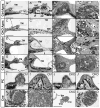
Figure 3. Conditional deletion of Atoh1 generates a flat epithelium and shows long-term retention of some spiral neurons in neonates
(AH′) Thin plastic sections through cochlea show histological changes of P14 CKO ear. The control mice form the spiral limbus and sulcus, the tectorial membrane and the organ of Corti on the basilar membrane (A, A′). Sections through the apex of the mutant cochlear reveal a flat epithelium consisting of a single row of cuboidal cells on the basilar membrane (C, C′). While the basilar membrane is progressively shorter toward the base, there is no alteration in the flat epithelium that indicates the position of the organ of Corti through the approximation of the tectorial membrane (E, E′, G, G′). Control mice have a large Rosenthal’s canal filled with sensory neurons (B, B′). In CKO mice the apex and the basal tip have clusters of sensory neurons (D, D′, H, H′), whereas the middle turn has just a bony matrix or a few isolated neurons surrounded by bone (arrows in F and F′). Note that all bone spaces are filled with neurons in both control and Atoh1 CKO mutants, suggesting that the neuronal cell death phase is not continuing in neonates despite the transformation of the primordial organ of Corti into a flat epithelium in the absence of Atoh1. (I–J′) Some hair cells are only found in the posterior canal crista in the mutant ear. Sections through the posterior canal crista of control mice show the large bundle of the posterior canal crista nerve approaching an organ filled with many hair cells that extend apical bundles into the cupula (I, I′). The nerve approaching the posterior canal crista of Atoh1 CKO mice are much smaller and some areas of the crista show no hair cells (J, J′, arrow indicates a differentiated hair cell). (K–L′) Sections through the cochlear nerve show obvious reduction of the number of nerve fibers in the CKO mutant in comparison to control (See Table 1). BM, basilar membrane; Cne, cochlear nerve; Eff, efferent nerve fibers; IHC, inner hair cell; IP, inner pillar cell; OHC, outer hair cell; OP, outer pillar cell; PC, posterior canal crista; PCN, posterior canal crista nerve; SG, spiral ganglion; SL, spiral limbus; TC, tunnel of Corti; TM, tectorial membrane; Vne, vestibular nerve. Bar indicates 100 μm in all except in A′, B′, C′, D′, E′, F′, G′, H′, K′ and L′, where it indicates 10 μm.
Figure 4
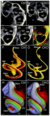
Figure 4. TSLIM reveals comparable cochlear development with near identical length of the cochlea in CKO mutant
(A–E) TSLIM 5 μm optical sections through the cochlea at comparable positions of P21 control and CKO mice show some reduction of the scalae and apparent radial reduction of basilar membrane. The narrowing of the mutant basilar membrane in the mutant ear is evident when the sections are aligned at the lateral end of the basilar membrane (arrow in E). (F–I) 3D reconstructions show that the overall structure of the CKO mutant cochlea is near normal despite the absence of an identifiable differentiated organ of Corti (bright red). The position of remaining Rosenthal’s canal (orange) in the mutant is consistent with the topology of neurons found in plastic sections shown in Fig. 3D′, F′, H′ (G). However, the volume reduction of scalae (scala media is made transparent, scala tympani in dark red and scala vestibule in light purple) , basilar membrane (yellow) and tectorial membrane (green) do not affect the longitudinal extension of the mutant cochlea (G, I). BM, basilar membrane; OC, organ of Corti; RC, Rosenthal’s canal; SM, scala media; ST, scala tympani; SV, scala vestibule; TM, tectorial membrane. Bar indicates 100 μm.
Figure 5
Figure 5. The pattern of residual innervation correlates with the distribution of Sox2 and Bdnf in Atoh1 CKO mice
(A–F) Immunolabeling with Tubulin shows the reduction of innervation in P0 Atoh1 CKO mice. Radial fibers are closely spaced in the cochlea of control mice (A). In contrast, the base of the Atoh1 CKO cochlea is almost free of any fibers (B) except for the basal tip (B′), whereas the apex (B″) and lower middle turn show some innervation with higher spacing density of radial fibers toward the apex (B, B″). The fibers in the middle turn demonstrate abnormal loop formation near the greater epithelial ridge (B). The vestibular innervation is present in all sensory epithelia but greatly reduced (D, F) in comparison to control littermate (C, E). (G–L″) Expression of Sox2 persists but is reduced in mutant cochlea. Sox2 in situ hybridization reveals a discontinuous or patchy distribution throughout the mutant cochlea (I, J, J′). Combining Tubulin with Sox2 immunochemistry reveals Sox2 labeling in supporting cells and prosensory cells with dense innervation in control animals (K–K″). In some CKO mutants Sox2 protein is more restricted and patchy in the apex (L). Nerve fibers are attracted to those patches and branch and end freely (L″). (M–N′) Bdnf expression is greatly reduced in the Atoh1 CKO cochlea. In situ hybridization shows an apex to base gradient of Bdnf expression in the newborn control mice (M, M′), whereas in the mutant ear, Bdnf expression is patchy in the apical half and is lost in the base (N, N′). AC, anterior canal crista; GER, greater epithelial ridge; HC, horizontal canal crista; IGSB, intraganglionic spiral bundle; OC, organ of Corti; ‘OC’, putative organ of Corti in Atoh1 CKO; PC, posterior canal crista; RF, radial fibers; S, saccule; Sc, supporting cells; SG, spiral ganglion; U, utricle. Bar indicates 100 μm.
Figure 6
Figure 6. Loss of Atoh1 causes failure of organ of Corti supporting cell differentiation
(A–D) Expression of Prox1 is decreased in the mutant cochlea. In situ hybridization shows expression of Prox1 in the supporting cells in P0 control mice (A, A′). It is restricted to only two rows of patchy undifferentiated cells in the CKO cochlea with very weak expression in others (C, C′). In later stages (P7), immunochemistry with both anti-Prox1 and anti-Tubulin antibodies shows five rows of supporting cells labeled in the control cochlea (circles in B). In the CKO mutant littermate, there are no supporting cells detected by these markers in the unspecified organ of Corti (D). However, the topology of the organ of Corti seems to be retained and indicated by an above background expression of Tubulin (D). Nerve fibers show reduction of radial growth possibly due to the absence of differentiated organ of Corti (D). (E–H) Sox2 immunochemistry also shows no labeling of supporting cells in the P7 CKO cochlea. Sox2 is expressed in both supporting cells (circles in E′) and cells in greater epithelial ridge (GER) in control cochlear, while in the CKO mutant, only the cells in GER are labeled with Sox2 (F–F″). The pattern of innervation in P7 CKO mutants forms loops before reaching the GER with occasional overshooting branches (arrows in F″ and G′) near middle turn. The base has even fewer nerve fibers (H). Cl, Claudius cells; GER, greater epithelial ridge; He, Hensen’s cells; Mid, the middle part of the cochlea; OC, organ of Corti; ‘OC’, putative organ of Corti in Atoh1 CKO; Sc, supporting cells; SG, spiral ganglion. Bar indicates 100 μm.
Figure 7
Figure 7. Some Myo7a positive cells develop later in postnatal CKO mice
Consistent with the absence of Atoh1 in situ signal in the Atoh1 CKO mice, no Myo7a immunopositive cells are found in the mutant embryos, newborns, or in most postnatal mice up to P7 (B). However in some CKO cochlea, especially in late stages, there are few Myo7a positive cells detected (C, E). These few Myo7a positive cells are seen either individually or in small clusters. They receive the majority of the remaining nerve fibers and are more laterally flanked by Tubulin positive cells shaped either like pillar cells or Deiter’s cells with thin phalangeal processes (arrows in C and E). IHC, inner hair cell; Mid, the middle part of the cochlea; OHC, outer hair cell; P, pillar cells; RF, radial fibers. Bar indicates 100 μm in A–C and 10 μm in D and E.
Figure 8
Figure 8. Expression of several genes defines the undifferentiated organ of Corti in Atoh1 CKO mutant
(A–D″) Bmp4 defines the lateral boundary of the organ of Corti and is retained in the apex of the mutant cochlea. Bmp4 in situ hybridization shows an apex to base gradient in Hensen’s and Claudius cells lateral to the hair cells of the organ of Corti in P0 and P7 control mice (A, A′, C–C″). Likewise, Atoh1 CKO mice display a gradient of Bmp4 expression that is almost restricted to the apical half (B, B′, D–D″) and progressively lost in the base of the cochlea from P0 to P7 (B, D). Note the wider Bmp4 expression area in D″ is more closer to the apex than the area in B’, both of which appear to be restricted to the Hensen’s cells and Claudius cells in the absence of differentiation of the organ of Corti (B′, D″). (E–H″) Fgf10 defines the medial boundary of the organ of Corti. Fgf10 is expressed in the greater epithelial ridge (GER) in both control animals (E, G–G″) and also in CKO mice medial to the undifferentiated organ of Corti (F, H–H″). Like Bmp4, Fgf10 is also downregulated in the basal half of cochlea in the absence of Atoh1 compared to control littermate. (I–N) Gata3 is expressed in the organ of Corti in P7 and P21 control mice and shows a similar expression pattern in the area of the putative organ of Corti in Atoh1 CKO mice. Higher magnifications and sections reveal the distribution of Gata3 in situ reaction product in the organ of Corti and adjacent cells in control mice (I′, M). In Atoh1 CKO mice, Gata3 is expressed in undifferentiated cells that are topographically equal to the organ of Corti cells (J′, N). Cl, Claudius cells; GER, greater epithelial ridge; He, Hensen’s cells; IHC, inner hair cell; OC, organ of Corti; ‘OC’, putative organ of Corti in Atoh1 CKO; OHC, outer hair cell; SG, spiral ganglion. Bar indicates 100 μm in all except in A′, B′, M, N where it indicates 10 μm.
Figure 9
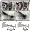
Figure 9. Flanking markers define the cellular loss of the organ of Corti in the Atoh1 CKO mutant
(A–B″) Fgf10 and Bmp4 expression shown by double in situ hybridization defines the region between the two expression domains to be the organ of Corti. The P1 control mice have differentiated supporting and hair cells in this region (A, A′, with I, 1, 2, 3 in A′ indicating the inner and three rows of outer hair cells respectively), whereas only two rows of undifferentiated cells remain in the Atoh1 CKO mice (B, B′). Radial sections show the degree of differentiation of hair cells and supporting cells (stars in A″) in the control cochlea between the two areas of marker expression (Fgf10 medially and Bmp4 laterally). At this stage, Fgf10 also appears to be expressed in the inner hair cells (arrow in A″). However in the mutant cochlea, the two areas of marker expression are only separated by two rows of cells of the putative organ of Corti (B″), with patchy cells at the position of inner hair cells also expressing Fgf10 (arrows in B′ and B″). (C, D) Schematic diagrams summarize the cellular loss of organ of Corti and the marker gene expression pattern change in the Atoh1 CKO mutant. Cl, Claudius cells; GER, greater epithelial ridge; He, Hensen’s cells; IHC, inner hair cell; LER, lesser epithelial ridge; Mid, the middle part of the cochlea; OC, organ of Corti; ‘OC’, putative organ of Corti in Atoh1 CKO; OHC, outer hair cells; SG, spiral ganglion; SL, spiral limbus. Bar indicates 100 μm in A, B and 10 μm in A′, A″, B′, B″.

Similar articles

Cited by

References

    1. Anniko M, Arnesen AR. Cochlear nerve topography and fiber spectrum in the pigmented mouse. Arch Otorhinolaryngol. 1988;245:155–9. - PubMed
    1. Bermingham-McDonogh O, Oesterle EC, Stone JS, Hume CR, Huynh HM, Hayashi T. Expression of Prox1 during mouse cochlear development. J Comp Neurol. 2006;496:172–86. - PMC - PubMed
    1. Bermingham NA, Hassan BA, Wang VY, Fernandez M, Banfi S, Bellen HJ, Fritzsch B, Zoghbi HY. Proprioceptor pathway development is dependent on Math1. Neuron. 2001;30:411–22. - PubMed
    1. Bermingham NA, Hassan BA, Price SD, Vollrath MA, Ben-Arie N, Eatock RA, Bellen HJ, Lysakowski A, Zoghbi HY. Math1: an essential gene for the generation of inner ear hair cells. Science. 1999;284:1837–41. - PubMed
    1. Campbell JP, Henson MM. Olivocochlear neurons in the brainstem of the mouse. Hear Res. 1988;35:271–4. - PubMed

Publication types