Review
. 2011 Feb;10(2):187-98.
doi: 10.1016/S1474-4422(10)70277-5. Epub 2010 Dec 10.

Disrupted energy metabolism and neuronal circuit dysfunction in cognitive impairment and Alzheimer's disease

Affiliations
Review

Disrupted energy metabolism and neuronal circuit dysfunction in cognitive impairment and Alzheimer's disease

Dimitrios Kapogiannis et al. Lancet Neurol. 2011 Feb.

Erratum in

  • Lancet Neurol. 2011 Feb;10(2):115

Abstract

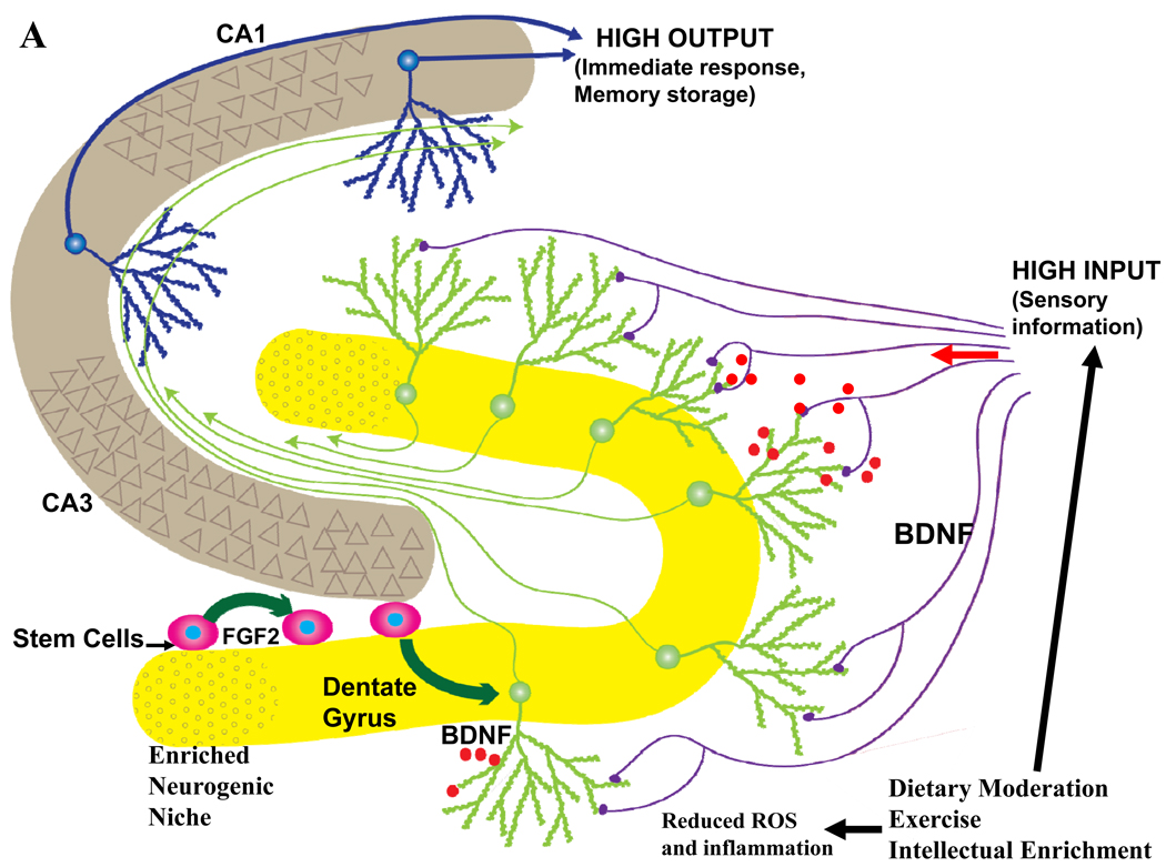
Epidemiological, neuropathological, and functional neuroimaging evidence implicates global and regional disruptions in brain metabolism and energetics in the pathogenesis of cognitive impairment. Nerve cell microcircuits are modified by excitatory and inhibitory synaptic activity and neurotrophic factors. Ageing and Alzheimer's disease cause perturbations in cellular energy metabolism, level of excitation or inhibition, and neurotrophic factor release, which overwhelm compensatory mechanisms and result in dysfunction of neuronal microcircuits and brain networks. A prolonged positive energy balance impairs the ability of neurons to adapt to oxidative and metabolic stress. Results from experimental studies in animals show how disruptions caused by chronic positive energy balance, such as diabetes, lead to accelerated cognitive ageing and Alzheimer's disease. Therapeutic interventions to allay cognitive dysfunction that target energy metabolism and adaptive stress responses (such as neurotrophin signalling) have been effective in animal models and in preliminary studies in humans.


Conflict of interest statement

Conflicts of Interest: Neither author has any conflicts of interest to declare.

Figures

Figure 1
Figure 1
Spread of the neuropathology in AD. Neurofibrillary tangles and neurodegeneration first appear in entorhinal cortex, and then in other medial temporal lobe (MTL) structures; fibrillary Aβ deposits and plaques first appear in transmodal areas [such as the posterior medial cortex (PMC), the inferior parietal lobule (IPL) and the lateral temporal lobe and temporal pole] that maintain reciprocal connections (illustrated by yellow arrows) with the entorhinal cortex. Spread of neurofibrillary tangles and neurodegeneration (illustrated by blue arrows) does not correlate with the spread of fibrillary Aβ deposition and plaque formation.
Figure 2
Figure 2
Evolution of cognitive ability with age, in the presence or absence of AD pathology: schematic progression of pathology, brain network dynamics and clinical manifestations. SCI: subjective cognitive impairment; MCI: mild cognitive impairment; AD: clinical Alzheimer’s disease; NFTs: neurofibrillary tangles; PMC: posterior medial cortex; MTL: medial temporal lobe.
Figure 3
Figure 3
Basic organization of neuronal microcircuits that control information flow through all brain regions involved in cognitive processing. The major excitatory projection neurons are glutamatergic with long axons that synapse on dendrites of other glutamatergic neurons that may, in turn, project their axons to a different brain region. GABAergic interneurons receive excitatory inputs from glutamatergic neurons and form synapses on the cell bodies of the same or other glutamatergic neurons. Glutamatergic neurons also receive synaptic inputs from noradrenergic, serotonergic and cholinergic neurons whose cell bodies are located in the locus ceruleus, raphe nucleus and basal forebrain, respectively. Neurons in all brain regions also interact with glial cells including astrocytes and microglia which produce trophic factors and cytokines which may normally play important roles in synaptic plasticity. However, excessive production of pro-inflammatory cytokines and reactive oxygen species (ROS) by glial cells has been implicated in the pathogenesis of cognitive impairment and AD.
Figure 4
Figure 4
Mechanisms of synaptic dysfunction in aging and Alzheimer’s disease. The β-amyloid precursor protein is axonally transported and so is present in high amounts in presynaptic terminals. In properly functioning synapses the APP is proteolytically cleaved in the middle of the Aβ sequence by the α-secretase, thereby preventing the production of Aβ. During normal aging, and more so in AD, APP is cleaved at the N- and C-termini of Aβ by β-secretase and γ-secretase, respectively, resulting in the production and self-aggregation of Aβ. Aggregation of Aβ on the membrane generates ROS resulting in membrane lipid peroxidation, which then impairs the function of membrane ion-motive ATPases thereby promoting membrane depolarization and Ca2+ influx through NMDA receptor channels and voltage-dependent Ca2+ channels. Sustained elevation of cytoplasmic Ca2+ levels promotes depletion of presynaptic glutamate stores resulting in impaired synaptic transmission and damage to axons and dendrites. In addition, perturbed mitochondrial function caused by aging, oxidative stress and Aβ results in energy depletion in neurons which exacerbates synaptic dysfunction and degeneration of neurons. Further contributing to the demise of neurons in AD is dysregulation of endoplasmic reticulum (ER) function that results in depletion of ER Ca2+ stores and accumulation of misfolded proteins.
Figure 5
Figure 5
The impact of lifelong ‘brain healthy’ and unhealthy lifestyles on late life hippocampal plasticity and cognitive function. Information from multimodal sensory association cortices enters the hippocampus from the entorhinal cortex via perforant path axons which synapse on dendrites of dentate granule neurons. The axons of granule neurons synapse on dendrites of pyramidal neurons which, in turn, may synapse on additional pyramidal projection neurons which then exit the hippocampus and innervate neurons in regions of the cerebral cortex involved in the long-term storage and processing of memories. A. Behaviors believed to promote healthy brain aging include moderation of dietary energy intake, regular exercise and engaging in intellectually challenging occupations and hobbies. Data suggest that these behaviors increase activity in hippocampal circuits and impose a mild cellular stress on neurons resulting in the activation of signaling pathways that induce the production of neurotrophic factors such as BDNF. As a consequence, synaptic plasticity and neurogenesis are enhanced and the resistance of neurons to aging and disease processes is increased. B. Behaviors that may contribute to cognitive impairment include excessive dietary energy intake, a sedentary lifestyle and a low level of cognitively challenging experiences. The latter lifestyle promotes diabetes and obesity, and can impair hippocampal synaptic plasticity and neurogenesis, thereby rendering neurons vulnerable to dysfunction and degeneration during aging.
Figure 5
Figure 5
The impact of lifelong ‘brain healthy’ and unhealthy lifestyles on late life hippocampal plasticity and cognitive function. Information from multimodal sensory association cortices enters the hippocampus from the entorhinal cortex via perforant path axons which synapse on dendrites of dentate granule neurons. The axons of granule neurons synapse on dendrites of pyramidal neurons which, in turn, may synapse on additional pyramidal projection neurons which then exit the hippocampus and innervate neurons in regions of the cerebral cortex involved in the long-term storage and processing of memories. A. Behaviors believed to promote healthy brain aging include moderation of dietary energy intake, regular exercise and engaging in intellectually challenging occupations and hobbies. Data suggest that these behaviors increase activity in hippocampal circuits and impose a mild cellular stress on neurons resulting in the activation of signaling pathways that induce the production of neurotrophic factors such as BDNF. As a consequence, synaptic plasticity and neurogenesis are enhanced and the resistance of neurons to aging and disease processes is increased. B. Behaviors that may contribute to cognitive impairment include excessive dietary energy intake, a sedentary lifestyle and a low level of cognitively challenging experiences. The latter lifestyle promotes diabetes and obesity, and can impair hippocampal synaptic plasticity and neurogenesis, thereby rendering neurons vulnerable to dysfunction and degeneration during aging.

References

    1. Gunstad J, Lhotsky A, Wendell CR, et al. Longitudinal Examination of Obesity and Cognitive Function: Results from the Baltimore Longitudinal Study of Aging. Neuroepidemiology. 2010;34:222–2229. - PMC - PubMed
    1. Luchsinger JA, Reitz C, Patel B, et al. Relation of diabetes to mild cognitive impairment. Archives of neurology. 2007;64:570–575. - PubMed
    1. Toro P, Schonknecht P, Schroder J. Type II diabetes in mild cognitive impairment and Alzheimer's disease: results from a prospective population-based study in Germany. J Alzheimers Dis. 2009;16:687–691. - PubMed
    1. Sperling RA, Dickerson BC, Pihlajamaki M, et al. Functional alterations in memory networks in early Alzheimer's disease. Neuromolecular Med. 2010;12:27–43. - PMC - PubMed
    1. Fouquet M, Desgranges B, Landeau B, et al. Longitudinal brain metabolic changes from amnestic mild cognitive impairment to Alzheimer's disease. Brain. 2009;132:2058–2067. - PMC - PubMed

Publication types