. 2011 Feb 10:1373:240-52.
doi: 10.1016/j.brainres.2010.12.015. Epub 2010 Dec 11.

Differential response of the central noradrenergic nervous system to the loss of locus coeruleus neurons in Parkinson's disease and Alzheimer's disease

Affiliations

Differential response of the central noradrenergic nervous system to the loss of locus coeruleus neurons in Parkinson's disease and Alzheimer's disease

Pamela J McMillan et al. Brain Res. .

Abstract

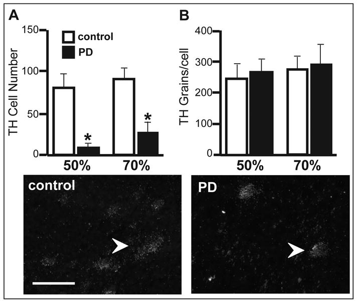
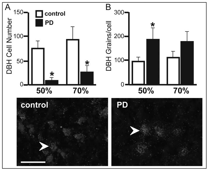
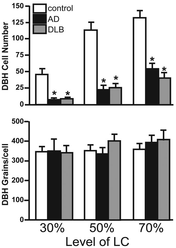
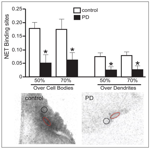
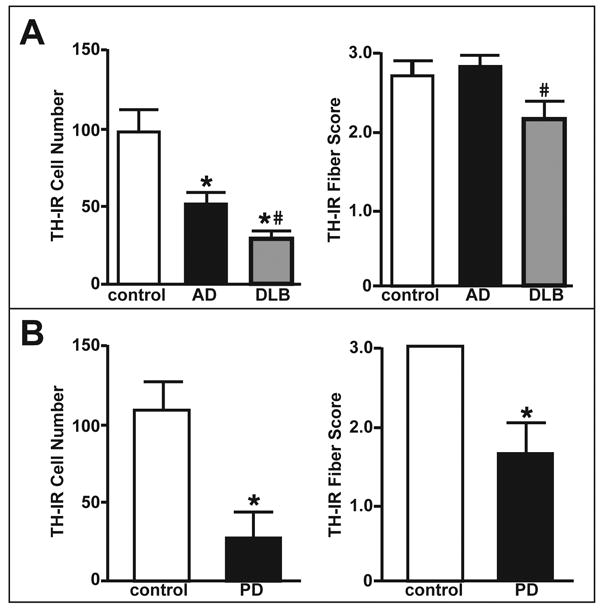
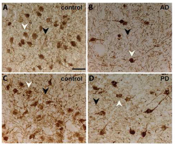
In Parkinson's disease (PD), there is a significant loss of noradrenergic neurons in the locus coeruleus (LC) in addition to the loss of dopaminergic neurons in the substantia nigra (SN). The goal of this study was to determine if the surviving LC noradrenergic neurons in PD demonstrate compensatory changes in response to the neuronal loss, as observed in Alzheimer's disease (AD). Tyrosine hydroxylase (TH) and dopamine β-hydroxylase (DBH) mRNA expression in postmortem LC tissue of control and age-matched PD subjects demonstrated a significant reduction in the number of noradrenergic neurons in the LC of PD subjects. TH mRNA expression/neuron did not differ between control and PD subjects, but DBH mRNA expression/neuron was significantly elevated in PD subjects compared to control. This increase in DBH mRNA expression in PD subjects is not a response to neuronal loss because the amount of DBH mRNA expression/neuron in AD subjects was not significantly different from control. Norepinephrine transporter (NET) binding site concentration in the LC of PD subjects was significantly reduced over the cell body region as well as the peri-LC dendritic zone. In PD subjects, the loss of dendrites from surviving noradrenergic neurons was also apparent with TH-immunoreactivity (IR). This loss of LC dendritic innervation in PD subjects as measured by TH-IR was not due to LC neuronal loss because TH-IR in AD subjects was robust, despite a similar loss of LC neurons. These data suggest that there is a differential response of the noradrenergic nervous system in PD compared to AD in response to the loss of LC neurons.


Figures

Figure 1
Figure 1
TH mRNA expression in the LC of age-matched control (n=8) and PD (n=7) subjects. (A) The number of TH positively labeled neurons at the 50 and 70% level of the LC of age-matched control and PD subjects. In PD subjects, there is a significant decrease in the number of TH positively labeled neurons at both levels of the LC (50% level p=0.009, 70% level p=0.008). (B) Expression of TH mRNA/neuron in the LC of control and PD subjects at the 50 and 70% levels. All labeled neurons that were counted as positively labeled were also quantitated for the amount of TH mRNA expression/neuron. There is not a difference in the amount of TH mRNA expression/neuron in PD subjects as compared to control age-matched subjects. The bottom two images are dark-field photomicrographs of TH mRNA labeled neurons in age-matched control (bottom left) and PD (bottom right) at the 50% level of LC. * Indicates significant difference from age-matched control subjects. Arrow indicates a TH mRNA labeled neuron. Scale bar = 100μm.
Figure 2
Figure 2
DBH mRNA expression in the LC of age-matched control (n=8) and PD (n=7) subjects. (A) The number of DBH positively labeled neurons at the 50 and 70% level of the LC of age-matched control and PD subjects. In PD subjects, there is a significant decrease in the number of DBH positively labeled neurons at both levels of the LC (50% level p=0.007, 70% level p=0.03). (B) Expression of DBH mRNA/neuron in the LC of age-matched control and PD subjects at the 50 and 70% levels. All labeled neurons that were counted as positively labeled were also quantitated for the amount of DBH mRNA expression/neuron. There is a significant increase in the amount of DBH mRNA expression/neuron at the 50% (p=0.03) level of the LC in PD subjects, as compared to control age-matched subjects. The bottom two images are dark-field photomicrographs of DBH mRNA labeled neurons in age-matched control (bottom left) and PD (bottom right) at the 50% level of LC. * Indicates significant difference from age-matched control subjects. Arrow indicates a DBH mRNA labeled neuron. Scale bar = 100μm.
Figure 3
Figure 3
DBH mRNA expression in the LC of age-matched control (n=15), AD (n=15) and DLB (n=13) subjects. (A) The number of DBH positively labeled neurons at the 30 (F(2,38,40) =17.1, p<0.0001), 50 (F(2,37,39) =38.0, p<0.0001) and 70% (F(2,39,41) =29.7, p<0.0001) level of the LC of age-matched control, AD, and DLB subjects. In AD and DLB subjects, there is a significant decrease in the number of DBH positively labeled neurons at all three levels of the LC. (B) Expression of DBH mRNA/neuron in the LC of age-matched control, AD, and DLB subjects at the 30, 50 and 70% levels. All labeled neurons that were counted as positively labeled were also quantitated for the amount of DBH mRNA expression/neuron. There is not a difference in the amount of DBH mRNA expression/neuron in the surviving LC neurons in AD and DLB subjects as compared to control age-matched subjects. * Indicates significant difference from age-matched control subjects.
Figure 4
Figure 4
NET binding site concentrations over the LC region in age-matched control (n=8) and PD (n=7) subjects. Top panel: NET binding site concentrations over LC cell body region and over the peri-LC dendritic zone at the 50 and 70% level of the LC in age-matched control and PD subjects. NET binding site concentrations are significantly reduced in PD subjects at both levels of the LC and in both regions of the LC as compared to age-matched control subjects (cell body region 50% level p=0.006, 70% level p=0.03; dendritic region 50% level p=0.012, 70% level p=0.015). Bottom panel: Autoradiographic image of NET in the LC of age-matched control and PD subjects. * Indicates significant difference from age-matched control subjects. The dark circle represents NET binding over the peri-LC dendritic zone and the grey oblong represents NET binding over LC body region.
Figure 5
Figure 5
Comparison of TH-IR at the 70% level of the LC of control, AD/PD and PD subjects. (A) The number of TH-IR positive neurons (left histogram) and the density of TH-IR fibers (right histogram) in control, AD and DLB subjects. The number of TH-IR neurons is significantly decreased in AD and DLB subjects as compared to control (F(2,32,34) =11.8, p=0.0001), while TH-IR fibers in AD and DLB subjects are not significantly different from controls. DLB subjects exhibit a further reduction as compared to AD subjects. (B) The number of TH-IR positive neurons (left histogram) and the density of TH-IR fibers (right histogram) in control and PD subjects. The number of TH-IR neurons (p=0.008) and TH-IR fiber (p=0.004) density are significantly decreased in PD subjects as compared to control subjects. * Indicates significant difference from age-matched control subjects. # Indicates significant difference from AD subjects.
Figure 6
Figure 6
Photomicrographs of TH-IR labeling in the LC at the 70% level in control (A and C), AD (B) and PD (D) subjects. White arrowhead indicates TH-IR labeled cell body and dark arrowhead indicates a TH-IR labeled fiber tract. Scale Bar = 100 μm.

Similar articles

Cited by

References

    1. Adolfsson R, Gottfries CG, Roos BE, Winblad B. Changes in the brain catecholamines in patients with dementia of Alzheimer's type. Br J Psychiatry. 1979;135:216–223. - PubMed
    1. Arai A, Tomiyama M, Kannari K, Kimura T, Suzuki C, Watanabe M, Kawarabayashi T, Shen H, Shoji M. Reuptake of L-DOPA-derived extracellular DA in the striatum of a rodent model of Parkinson's disease via norepinephrine transporter. Synapse. 2008;62:632–635. - PubMed
    1. Aston-Jones G, Zhu Y, Card JP. Numerous GABAergic afferents to locus cerleus in the pericerulear dendritic zone: possible interneuronal pool. J Neurosci. 2004;24:2313–2321. - PMC - PubMed
    1. Ballard C, Holmes C, McKeith I, Neill D, O'Brien J, Cairns N, Lantos P, Perry E, Ince P, Perry R. Psychiatric morbidity in dementia with Lewy bodies: a prospective clinical and neuropathological comparative study with Alzheimer's disease. Am J Psychiatry. 1999;156:1039–1045. - PubMed
    1. Barber R, Panikkar A, McKeith IG. Dementia with Lewy bodies: diagnosis and management. Int J Geriatr Psychiatry. 2001;16:S12–S18. - PubMed

Publication types

MeSH terms