. 2011 May 30;191(1-3):288-95.
doi: 10.1016/j.cbi.2010.12.002. Epub 2010 Dec 11.

Bioenergetic function in cardiovascular cells: the importance of the reserve capacity and its biological regulation

Affiliations

Bioenergetic function in cardiovascular cells: the importance of the reserve capacity and its biological regulation

Brian E Sansbury et al. Chem Biol Interact. .

Abstract

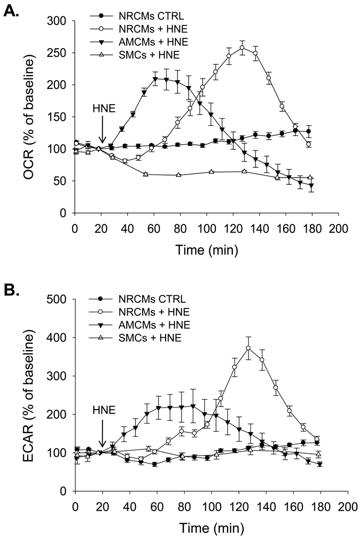
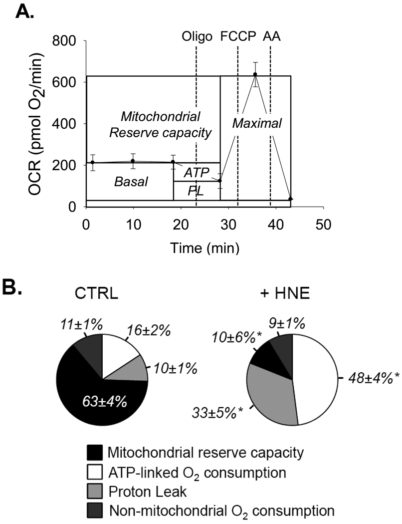
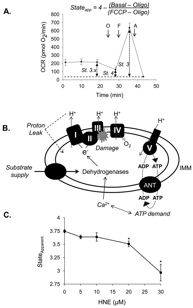
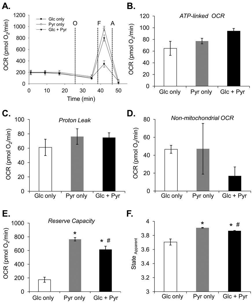
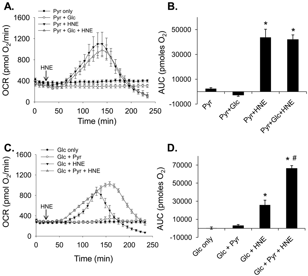
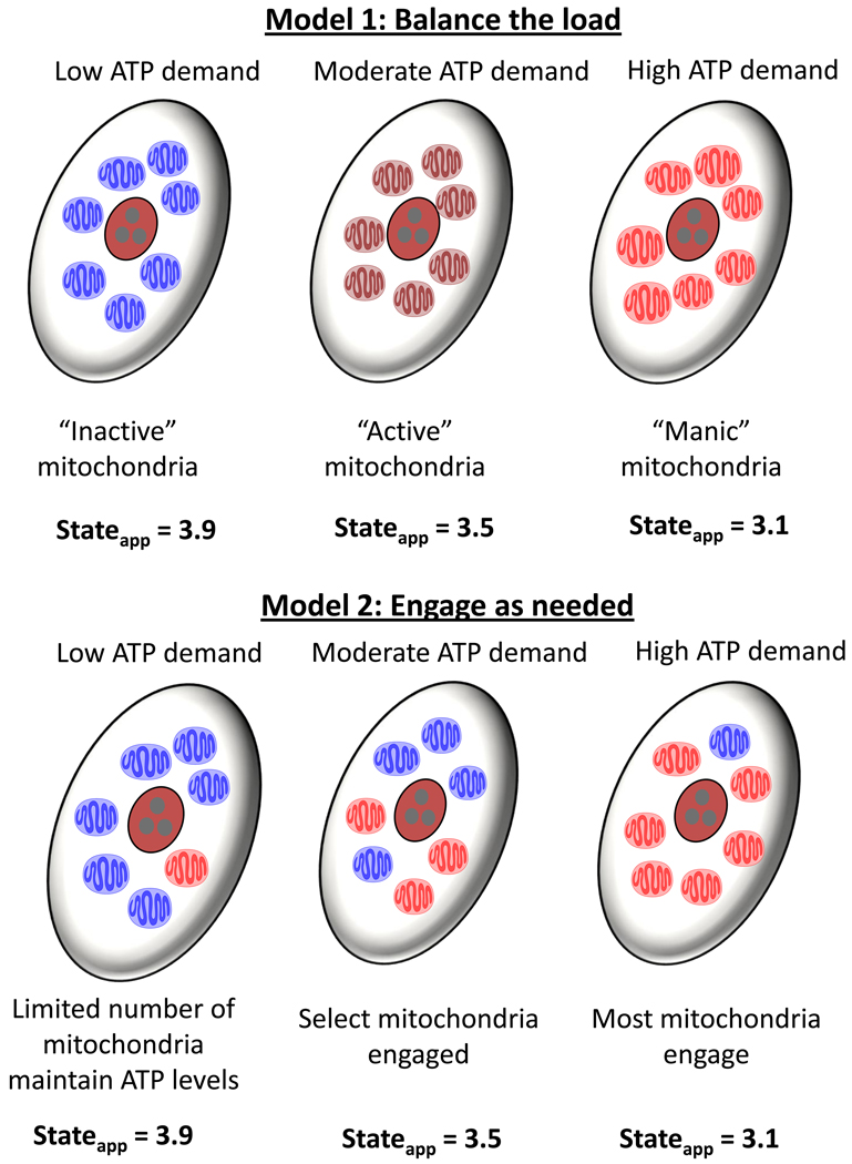
The ability of the cell to generate sufficient energy through oxidative phosphorylation and to maintain healthy pools of mitochondria are critical for survival and maintenance of normal biological function, especially during periods of increased oxidative stress. Mitochondria in most cardiovascular cells function at a basal level that only draws upon a small fraction of the total bioenergetic capability of the organelle; the apparent respiratory state of mitochondria in these cells is often close to state 4. The difference between the basal and maximal activity, equivalent to state 3, of the respiratory chain is called the reserve capacity. We hypothesize that the reserve capacity serves the increased energy demands for maintenance of organ function and cellular repair. However, the factors that determine the volume of the reserve capacity and its relevance to biology are not well understood. In this study, we first examined whether responses to 4-hydroxynonenal (HNE), a lipid peroxidation product found in atherosclerotic lesions and the diseased heart, differ between vascular smooth muscle cells, adult mouse cardiomyocytes, and rat neonatal cardiomyocytes. In both types of cardiomyocytes, oxygen consumption increased after HNE treatment, while oxygen consumption in smooth muscle cells decreased. The increase in oxygen consumption in cardiomyocytes decreased the reserve capacity and shifted the apparent respiratory state closer to state 3. Neonatal rat cardiomyocytes respiring on pyruvate alone had a fourfold higher reserve capacity than cells with glucose as the sole substrate, and these cells were more resistant to mitochondrial dysfunction induced by 4-HNE. The integration of the concepts of reserve capacity and state-apparent are discussed along with the proposal of two potential models by which mitochondria respond to stress.


Figures

Figure 1
Figure 1. Bioenergetic responses of different cardiovascular cells to electrophile stress
Extracellular flux analysis of oxygen consumption and glycolysis in neonatal rat cardiomyocytes (NRCMs), adult mouse cardiomyocytes (AMCMs), and rat aortic smooth muscle cells (SMCs). Three baseline measurements of oxygen consumption rate (OCR; panel A) or extracellular acidification rate (ECAR; panel B) were recorded from isolated cardiomyocytes or SMC. Vehicle (DMEM) or HNE was then injected to a final concentration of 20 µM and the measurements then continued for the indicated times. n = 3–5 per group; Repeated measure one-way ANOVA indicated that the response of the groups in panels A and B to HNE differed with respect to time (p<0.05).
Figure 2
Figure 2. Measurement of bioenergetic parameters in myocytes using XF technology
(A) Mitochondrial function assay in primary cardiomyocytes: After three baseline OCR measurements, oligomycin (1 µg/ml), FCCP (1 µM) and antimycin A (10 µM) were injected sequentially. OCR measurements were recorded after each injection. ATP-linked oxygen consumption (ATP) and the OCR due to proton leak (PL) can be calculated using the basal OCR rate and the oligomycin-insensitive rate. Injection of the uncoupling agent FCCP is used to determine the maximal respiratory capacity, and injection of antimycin A allows for the measurement of non-mitochondrial oxygen consumption. The reserve capacity is calculated by subtracting the maximal rate of oxygen consumption by the basal OCR. (B) Pie charts illustrating the effects of HNE on parameters of mitochondrial function. After three baseline rates, HNE was injected to 10 µM. After approximately 2h, oligomycin, FCCP, and antimycin A were injected sequentially and parameters of mitochondrial function were calculated as described in panel A. n = 3–4 per group, *p<0.05.
Figure 3
Figure 3. Effects of electrophile stress on the mitochondrial respiratory stateapparent in cardiomyocytes
Defining the mitochondrial respiratory stateapparent using the mitochondrial function assay: (A) After three baseline oxygen consumption rate (OCR) measurements, oligomycin (1 µg/ml), FCCP (1 µM) and antimycin A (10 µM) are injected sequentially, and OCR measurements are recorded after each injection. The basal rate of oxygen consumption describes the average respiratory state of mitochondria in the cell, which is described here as state 3.x. Exposure of cells to oligomycin puts mitochondria in state 4, and FCCP is then used to increase the respiration rate to a maximal rate comparable to state 3. The equation shown above the figure can then be used to calculate the respiratory stateapparent, where Basal represents the basal OCR, Oligo represents the OCR after oligomycin exposure, and FCCP represents the FCCP-stimulated OCR. The non-mitochondrial rate of oxygen consumption should be subtracted from all values prior to the calculation. (B) Schematic illustrating the modules that regulate reserve capacity and mitochondrial state apparent. Factors regulating respiration rate are italicized. (C) Effects of HNE on the respiratory stateapparent in cardiomyocytes: NRCMs were treated with HNE at the concentrations indicated and subjected to the energetic assay shown in panel A. n = 3–4 per group, *p<0.05.
Figure 4
Figure 4. Substrate-dependent changes in mitochondrial function
Extracellular flux analysis of cardiomyocytes in medium containing pyruvate and/or glucose: (A) Bioenergetic function assay in cardiomyocytes having glucose (Glc only), pyruvate (Pyr only), or glucose and pyruvate (Glc + Pyr) as substrate. ATP-linked OCR (B), proton leak (C), non-mitochondrial OCR (D), reserve capacity (E), and the mitochondrial respiratory stateapparent (F) were calculated from the assay in panel A. n = 5 per group, *p<0.05 vs. Glc only group; #p<0.05 vs Pyr only group.
Figure 5
Figure 5. Effects of substrate on the bioenergetic response of cardiomyocytes to HNE
Extracellular flux analysis of cardiomyocytes exposed to HNE in the presence of glucose and/or pyruvate. (A) One hour prior to energetic assays, the culture medium was removed and changed to medium containing pyruvate alone (1 mM; Pyr only) or pyruvate in the presence of glucose (5.5 mM; Pyr + Glc). After three baseline OCR measurements, HNE was injected to a final concentration of 20 µM in one of the pyruvate only groups (Pyr + HNE) and in one of the groups containing both pyruvate and glucose (Pyr + Glc + HNE), and rates were recorded for the indicated time. (B) Area under the curve (AUC) analysis of oxygen consumption rates from panel A: After baseline measurements, the AUC was measured to indicate the total amount of oxygen consumed in each group. (C) Cells were incubated in medium containing glucose alone (5.5 mM; Glc only) or glucose in the presence of pyruvate (Glc + Pyr). After three baseline OCR measurements, HNE was injected to a final concentration of 20 µM in one of the Glc only groups (Glc + HNE) and in one of the groups containing both glucose and pyruvate (Glc + Pyr + HNE), and rates were recorded for the indicated time. (D) Area under the curve (AUC) analysis of oxygen consumption rates from panel C: After baseline measurements, the AUC was measured to indicate the total amount of oxygen consumed in each group. n = 5 per group, *p<0.05 vs. cells not treated with HNE; #p<0.05 vs. Glc + HNE group.
Figure 6
Figure 6. Proposed models of mitochondrial responses to stress
Two simple models by which mitochondria respond to insults that increase ATP demand. Model 1, “Balance the load” model: When the need for energy arises, mitochondria respond collectively to increase ATP. A moderate demand is matched by a commensurate change in the mitochondrial stateapparent to somewhere in the midrange (e.g., state 3.5). Upon a strong challenge, all mitochondria in the cell respond and increase respiration to near their maximal rate (e.g., state 3.1). Model 2, “Engage when needed” model: In this model, most mitochondria in cells with low ATP demand idle at a respiratory state near 4, and only a subset of mitochondria work at a respiratory state more toward 3 to uphold basal energy requirements. When a moderate demand for energy arises, the cell then engages other subsets of mitochondria to generate the required amount of ATP. Under these conditions, some mitochondria might be near state 3.9 while others are more near state 3.1, averaging a collective stateapparent of 3.5. During times of very high energy demand, more mitochondria engage, yet a subpopulation of mitochondria remains in a resting state.

References

    1. Akao M, Ohler A, O'Rourke B, Marban E. Mitochondrial ATP-sensitive potassium channels inhibit apoptosis induced by oxidative stress in cardiac cells. Circ Res. 2001;88:1267–1275. - PubMed
    1. Aon MA, Cortassa S, O'Rourke B. Percolation and criticality in a mitochondrial network. Proc Natl Acad Sci U S A. 2004;101:4447–4452. - PMC - PubMed
    1. Aon MA, Cortassa S, O'Rourke B. The fundamental organization of cardiac mitochondria as a network of coupled oscillators. Biophys J. 2006;91:4317–4327. - PMC - PubMed
    1. Brand MD. The proton leak across the mitochondrial inner membrane. Biochim Biophys Acta. 1990;1018:128–133. - PubMed
    1. Brown GC. The leaks and slips of bioenergetic membranes. FASEB J. 1992;6:2961–2965. - PubMed

Publication types

LinkOut - more resources