. 2011 Mar;140(3):947-56.
doi: 10.1053/j.gastro.2010.12.005. Epub 2010 Dec 11.

IgA-producing plasma cells originate from germinal centers that are induced by B-cell receptor engagement in humans

Affiliations

IgA-producing plasma cells originate from germinal centers that are induced by B-cell receptor engagement in humans

Francesca Barone et al. Gastroenterology. 2011 Mar.

Abstract

Background & aims: IgA contributes to homeostatic balance between host and intestinal microbiota. Mechanisms that initiate the IgA response are unclear and likely to differ between humans and animal models. We used multiple experimental approaches to investigate the origin of human intestinal plasma cells that produce IgA in the gastrointestinal tract.

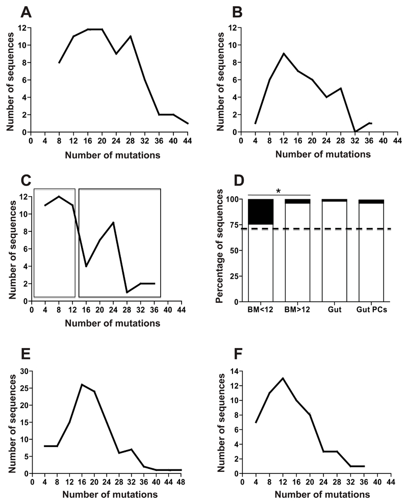
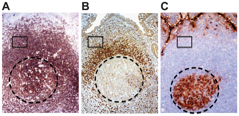
Methods: Complexity of IgA-producing plasma cell populations in human gastrointestinal mucosa and bone marrow and the specific response to oral cholera vaccine were compared by analysis of immunoglobulin genes. Flow cytometry, gene expression analysis, and immunohistochemistry were used to analyze signaling pathways induced by B-cell receptor engagement in human gut-associated lymphoid tissue (GALT) and involvement of innate immunity in B-cell activation in GALT compared with nonintestinal sites.

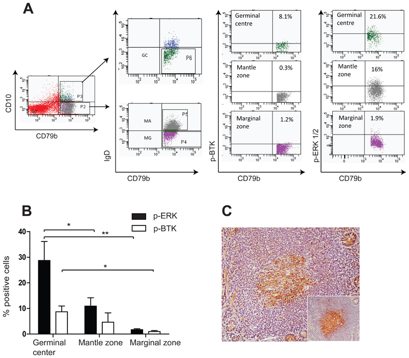
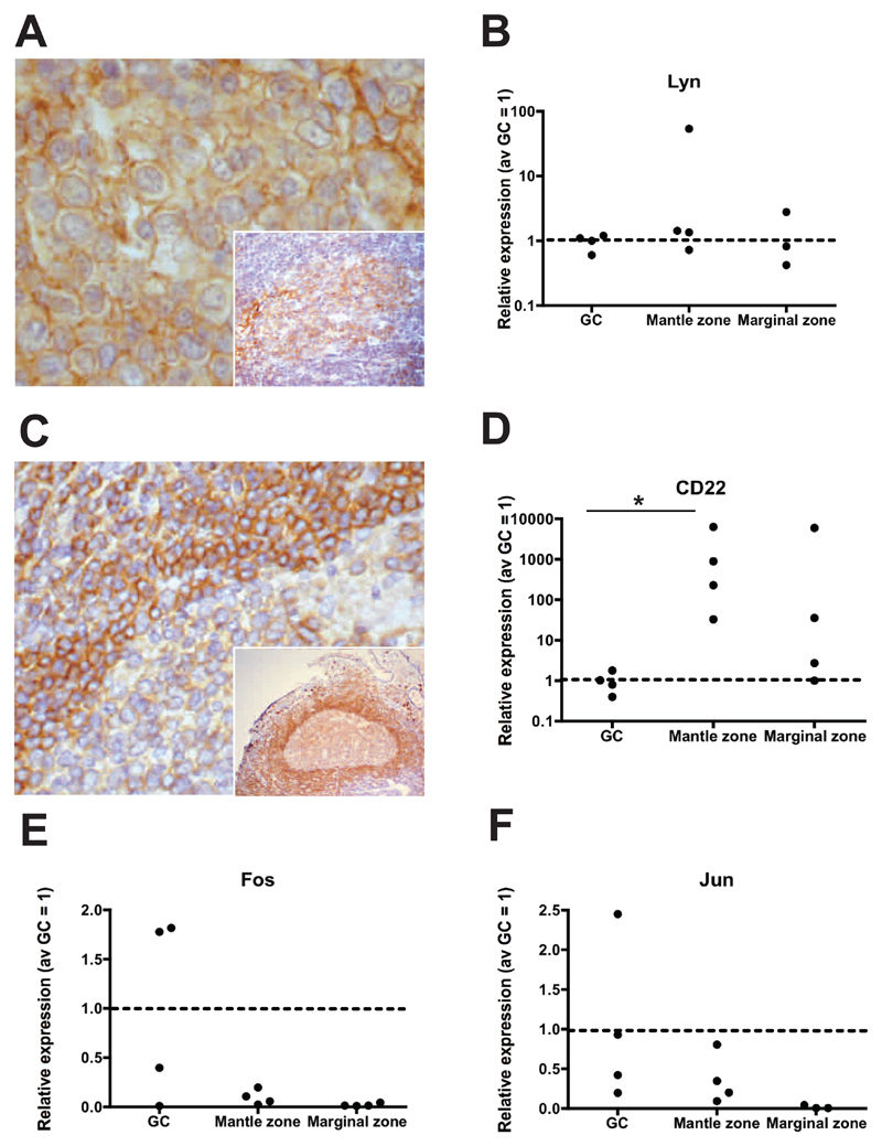
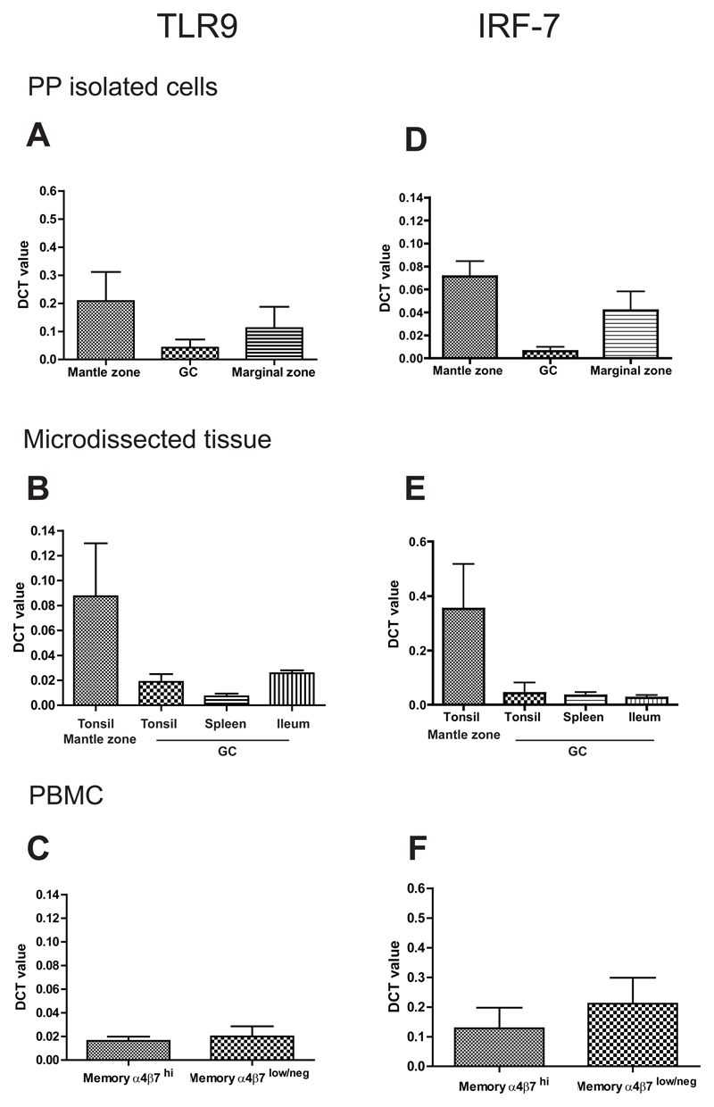
Results: Human intestinal IgA-producing plasma cells appeared to be of germinal center origin; there was no evidence for the population complexity that accompanies multiple pathways of derivation observed in bone marrow. In germinal center B cells of human GALT, Btk and Erk are phosphorylated, CD22 is down-regulated, Lyn is translocated to the cell membrane, and Fos and Jun are up-regulated; these features indicate B-cell receptor ligation during germinal center evolution. No differences in innate activation of B cells were observed in GALT, compared with peripheral immune compartments.

Conclusions: IgA-producing plasma cells appear to be derived from GALT germinal centers in humans. B-cell receptor engagement promotes formation of germinal centers of GALT, with no more evidence for innate immune receptor activation in the mucosa than nonintestinal immune compartments. Germinal centers in GALT should be targets of mucosal vaccinations because they are the source of human intestinal IgA response.


Conflict of interest statement

The authors declare that there is no conflict of interest.

Figures

Figure 1
Figure 1. Mucosal plasma cells comprise a single dominant population.
Charts illustrating the numbers of IGLV sequences (vertical axis) with different numbers of point mutations acquired by somatic hypermutation. Sequences are grouped on the horizontal axis as those with 0-4, 5-8, 9-12 etc mutations. Points represent the total number of sequences in the sampled population with the specified number of mutations. A. Mutations in rearrangements of IGLV1 and IGLV2 from plasma cells sampled from sections of ileal lamina propria from 3 individuals checked microscopically to be free of lymphoid tissue. B. Mutations in IGLV from single isolated colonic IgA+ plasma cells from 2 individuals. The distribution of mutations illustrated in A and B approximates to a single curve. C. Mutations in IGLV1 and IGLV2 used by IgA plasma cells from bone marrow from 3 individuals show 2 peaks in frequency of somatic hypermutation. D. Ratios of productive (white bar) to non-productive (black bar) rearrangements of IGLV1 and IGLV2. Sequences analysed were: BM<12 (IGLV1 and IGLV2 from bone marrow plasma cells with 12 mutations or fewer, as boxed in C), BM>12 (IGLV1 and IGLV2 from bone marrow plasma cells with more than 12 mutations as boxed in C), Gut (rearrangements of IGLV1 and IGLV2 from microdissected lamina propria cells shown in A) and Gut PCs (rearrangements of IGLV1 and IGLV2 from single isolated lamina propria plasma cells in B). Expected 71% in-frame is indicated with a dotted line. The ratio of in-frame: out of frame rearrangements of IGLV1 and IGLV2 in bone marrow plasma cells is not biased away from expected. However, there is a significant bias away from expected 71% in-frame rearrangements in the more mutated sequences from bone marrow, Gut and Gut PC groups (p=0.02, p<0.001 and p=0.01 respectively) and a significant difference between the more and less mutated subsets of IGLV1 and IGLV2 bone marrow plasma cells (p=0.03). E. Frequency of mutations in IGHV from blood-borne IgA+, α4β7hi cells from 3 individuals F. Mutations in IGHV from GC derived, blood-borne IgA+, α4β7hi B cells that bind CTB after an oral vaccination of 3 individuals with vaccine containing CTB display similar features to those from pre-immune blood not selected for any specificity.
Figure 2
Figure 2. B cell microenvironments in the Peyer’s Patches.
Sequential sections of PPs illustrating the organization of B cell populations. A. CD20 (brown) is expressed by the majority of B cells in the PPs, B. naïve B cells in the mantle express IgD, while marginal zone B cells, representing the largest population of B cell in the PPs are IgD-. C. GC B cell express CD10, while both marginal zone and mantle zone are CD10-. Circle indicates the GC areas, while boxes illustrate an area in the CD20+IgD-CD10- marginal zone. Original magnification 100x.
Figure 3
Figure 3. Btk and Erk are phosphorylated in PP GC B cells.
A. Example of flow cytometry scatter plot on human PP B cells showing immunostaining with anti-phosphorylated Btk (p-Btk) and phosphorylated Erk (p-Erk). Subsets of isolated B cells (CD79+) were identified on the basis of CD10 and IgD expression. GC cells (IgDCD10+) showed increased expression of phosphorylated Btk and Erk compared with mantle zone (IgD+CD10-) and marginal zone (IgD-CD10-) B cells. B. Diagram illustrating summary of replicate experiments with cells from 4 different individuals, immunostained with the antiphospho antibodies, demonstrating significantly increased p-Erk expression (p=0.05 and p=0.03 for GC v.s mantle zone and GC v.s. marginal zone, mean +SD) and increased p-Btk expression in GC B cells. C. Photomicrograph illustrating p-Btk expression within PP GCs (Original magnification 100x).
Figure 4
Figure 4. Signature of BCR activation in PP GCs.
A. Photomicrograph illustrating Lyn translocation to the cell membrane, in keeping with Lyn activation upon BCR engagement by antigen (and inset higher magnification). B. Relative quantitation of Lyn gene expression (4 individuals studied) by isolated GC (IgD-CD10+), mantle zone (IgD+CD10-) and marginal zone (IgD-CD10-) cells. Data is represented as relative quantitation normalized to average GC=1 (red dotted line). B cells isolated from PPs show no significant difference in Lyn mRNA expression in the three populations. C. IHC on PP GC showing low protein expression in the PP GCs immunostained with anti-CD22 monoclonal antibody, as compared with the mantle or marginal zones (and inset lower magnification). D. Accordingly, significant down-regulation of CD22 transcription in PP GCs was observed (p=0.03 GC vs. mantle zone). (Original magnification 200x in A and C and 100x in inset). E and F. Isolated PP GC cells show increased transcription of the BCR regulated genes, Jun and Fos.
Figure 5
Figure 5. No difference in TLR9 or IRF-7 expression in the GCs of Peyer’s Patches compared to GC from other lymphoid tissues.
Relative quantitation (DCT) of mRNA expression levels for TLR9 (A, B, C) in A. B cell subsets isolated from PP (GC, mantle and marginal zone; n=9 individual donors),B. microdissected areas of tonsils (GC and mantle zone n= 5 different donors), PP GCs (n= 7 individual donors) and spleen GCs (one donor). C. isolated mature mucosal (IgD-CD27+α4β7hi) and non mucosal (IgD-CD27+aα4β7lo/-) cells (n= 6 individual donors), showing no significant up-regulation of TLR9 transcript in GC B cells isolated from PP or microdissected tissue or mucosal B cells. D. Relative quantitation (DCT) of mRNA for IRF-7 in the same subsets analyzed for TLR9 (n=6 individual donors for each subset analyzed) showing lack of induction of IRF-7 gene in GC cells isolated from PP, E. microdissected GCs and F. sorted blood mucosal memory cells.

References

    1. Johansen FE, Pekna M, Norderhaug IN, et al. Absence of epithelial immunoglobulin A transport, with increased mucosal leakiness, in polymeric immunoglobulin receptor/secretory component-deficient mice. J Exp Med. 1999;190:915–22. - PMC - PubMed
    1. Fagarasan S, Muramatsu M, Suzuki K, et al. Critical roles of activation-induced cytidine deaminase in the homeostasis of gut flora. Science. 2002;298:1424–7. - PubMed
    1. Braathen R, Sandvik A, Berntzen G, et al. Identification of a polymeric Ig receptor binding phage-displayed peptide that exploits epithelial transcytosis without dimeric IgA competition. J Biol Chem. 2006;281:7075–81. - PubMed
    1. Walker WA, Isselbacher KJ, Bloch KJ. Immunologic control of soluble protein absorption from the small intestine: a gut-surface phenomenon. Am J Clin Nutr. 1974;27:1434–40. - PubMed
    1. Hapfelmeier S, Lawson MA, Slack E, et al. Reversible microbial colonization of germ-free mice reveals the dynamics of IgA immune responses. Science. 2010;328:1705–9. - PMC - PubMed

Publication types

MeSH terms

Substances