Review
. 2011 Jul;106(1):289-99.
doi: 10.1016/j.pbiomolbio.2010.11.010. Epub 2010 Dec 13.

Regulation of the cardiomyocyte population in the developing heart

Affiliations
Review

Regulation of the cardiomyocyte population in the developing heart

Kent Thornburg et al. Prog Biophys Mol Biol. 2011 Jul.

Abstract

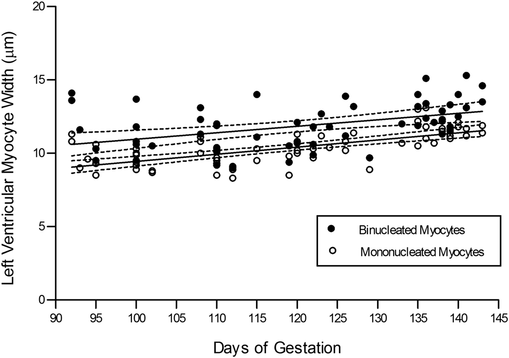
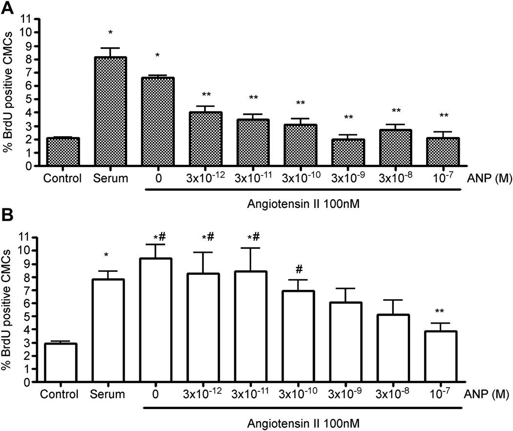
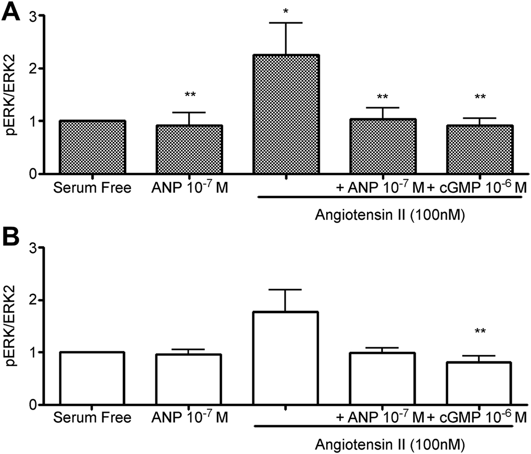
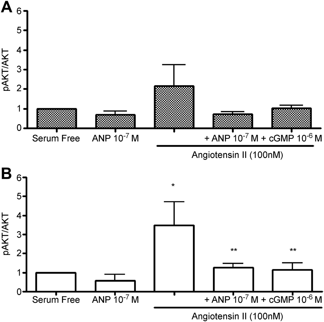
During fetal life the myocardium expands through replication of cardiomyocytes. In sheep, cardiomyocytes begin the process of becoming terminally differentiated at about 100 gestation days out of 145 days term. In this final step of development, cardiomyocytes become binucleated and stop dividing. The number of cells at birth is important in determining the number of cardiomyocytes for life. Therefore, the regulation of cardiomyocyte growth in the womb is critical to long term disease outcome. Growth factors that stimulate proliferation of fetal cardiomyocytes include angiotensin II, cortisol and insulin-like growth factor-1. Increased ventricular wall stress leads to short term increases in proliferation but longer-term loss of cardiomyocyte generative capacity. Two normally circulating hormones have been identified that suppress proliferation: atrial natriuretic peptide (ANP) and tri-iodo-L-thyronine (T₃). Atrial natriuretic peptide signals through the NPRA receptor that serves as a guanylate cyclase and signals through cGMP. ANP powerfully suppresses mitotic activity in cardiomyocytes in the presence of angiotensin II in culture. Addition of a cGMP analog has the same effect as ANP. ANP suppresses both the extracellular receptor kinases and the phosphoinositol-3 kinase pathways. T₃ also suppresses increased mitotic activity of stimulated cardiomyocytes but does so by increasing the cell cycle suppressant, p21, and decreasing the cell cycle activator, cyclin D1.


Figures

Figure 1
Figure 1
Cardiac myocytes from the fetal sheep with immuno-fluorescence staining for myosin (red; primary antibody Abcam ab15) and a Hoechst nuclear marker (cyan). Myocytes were also probed with an antibody against Ki-67 (DAB staining shown in yellow for contrast). The types of myocytes observed include, (A) a mononucleated myocyte positive for Ki-67 (arrow). (B) a cleavage furrow is clearly visible (arrowhead); (C) a dividing myocyte positive for Ki-67 (arrows) (D) a myocyte containing two nuclei that is positive for Ki-67 (arrow). Jonker, S.S. et al. J. Appl. Physiol. 2007. 102: 1130–1142.
Figure 2
Figure 2
Lengths of fetal sheep LV cardiac myocytes throughout the last third of gestation. The best-fit linear regression line and 95% confidence intervals of each data set are shown. Jonker, S et al. J. Appl. Physiol. 2007. 102: 1130–1142.
Figure 3
Figure 3
Widths of fetal sheep LV cardiac myocytes throughout the last third of gestation. The best-fit linear regression line and 95% confidence intervals of each data set are shown. Jonker, S.S. et al. J. Appl. Physiol. 2007. 102:1130–1142.
Figure 4
Figure 4
The proportion of total LV cardiac myocytes that are mononucleated during the last third of gestation. The proportion of mononucleated myocytes begins to decrease after ~100dGA. Most of these myocytes become binucleated. Shown are Boltzmann’s sigmoidal equations fit to each data set and the 95% confidence intervals. Jonker, S.S. et al. J. Appl. Physiol. 2007. 102: 1130–1142.
Figure 5
Figure 5
The proportion of all cardiac myocytes that are in the cell cycle decreases towards term. Inset panel: Cell cycle activity (Ki-67) normalized to the proportion of myocytes that are mononucleated and thus capable of proliferating or becoming terminally differentiated. The best-fit linear regression line and 95% confidence intervals are shown. Jonker, S.S. et al. J. Appl. Physiol. 2007. 102: 1130–1142.
Figure 6
Figure 6
The calculated total number of cardiac myocytes increased throughout the last third of gestation in the LV. The number of binucleated myocytes increased at the expense of mononucleated myocytes, which declined in number following ~115dGA. The proportion of myocytes becoming binucleated (rather than proliferating) increased rapidly after ~100dGA. Jonker, S.S. et al. J. Appl. Physiol. 2007. 102: 1130–1142.
Figure 7
Figure 7
IGF-1 receptor (IGF1R) signaling in cardiomyocytes. Isolated near-term fetal sheep cardiomyocytes were treated with the IGF-1 analog Long R3 IGF-1 (LR3 IGF-1, 1µg/ml), the PI3K inhibitor LY294002 (10 µM) and the MEK inhibitor UO126 (10 µM). Panels A and B show a representative Western blot for ERK (A) and AKT (B) stimulation. C and D: values are plotted as median fold change ± SE of blot density normalized to baseline LR3 IGF-1 stimulation (0 min, n=4). C: density is plotted in normalized units of phospho-ERK (p-ERK)/ERK2. D: density is plotted in normalized units of phospho-AKT (p-AKT)/AKT. *P<0.05 compared with within-treatment baseline. †P<0.05 compared with LR3 IGF-1 treatment at same time point. Sundgren, N.C. et al. Am. J. Physiol. Regul. Integr. Comp. Physiol. 2003. 285: R1481–R1489.
Figure 8
Figure 8
Ang II-stimulated BrdU uptake in cardiomyocytes isolated from the right (A) and left (B) fetal ventricles was inhibited by ANP in a dose-dependent manner (48h treatment). Left ventricular cardiomyocytes were less sensitive to ANP than were right ventricular cardiomyocytes. Data are means ± SEM. n=6 fetuses per group. #P<0.05 vs. matching treatment in RV; *P<0.05 vs. serum-free control in same ventricle; **P<0.05 vs. Ang II alone in same ventricle. O’Tierney, P.F. et al. Journal of Physiology, 2010. 588, 2879–2889.
Figure 9
Figure 9
Ang II-stimulated BrdU uptake in cardiomyocytes isolated from right (A) and left (B) fetal ventricles was inhibited by 48 hours of treatment with the cGMP analog, 8-bromo-cGMP (10−6M) and atrial natriuretic peptide (ANP, 10−7M). Control cells were cultured in serum-free conditions. Data are means ± SEM. n=5 or 6 fetuses per group. *P<0.05 vs. control; **P<0.05 vs. Ang II alone. O’Tierney, P.F. et al. Journal of Physiology, 2010. 588, 2879–2889.
Figure 10
Figure 10
ANP inhibits Ang II-induced ERK phosphorylation in fetal cardiomyocytes isolated from the right ventricle (A) with a similar trend for the left ventricle (B). cGMP suppresses the generation of phospho-ERK in right and left ventricular cardiomyocytes. Data are means ± SEM. n=5 or 6 fetuses per group. All treatments were 10 minutes. *P<0.05 vs. serum-free control; **P<0.05 vs. Ang II alone. O’Tierney, P.F. et al. Journal of Physiology, 2010. 588, 2879–2889.
Figure 11
Figure 11
The effects of ANP on Ang II-induced AKT phosphorylation in fetal cardiomyocytes isolated from the right (A) and left (B) ventricles. AKT phosphorylation was significantly increased in the presence of Ang II, an effect which was suppressed by ANP in the left ventricle (B) with a similar trend in the right ventricle (A). All treatments were 10 minutes. Data are means ± SEM. n=5 or 6 fetuses per group. *P<0.05 vs. serum-free controls; **P<0.05 vs. Ang II alone. O’Tierney, P.F. et al. Journal of Physiology, 2010. 588, 2879–2889.
Figure 12
Figure 12
BrdU (10uM) incorporation in isolated (A) LV and (B) RV fetal sheep cardiomyocytes with 10% FBS-serum media challenge. T3 at all doses (in serum media; S) significantly inhibited BrdU uptake. Data are means ± SEM. n=5 fetuses per group. *P<0.001 vs serum. Chattergoon, N.N. et al. J. Endocrinol. 2007. 192:R1–8.
Figure 13
Figure 13
Cyclin D1 and p21 expression (A, B respectively) in LV cardiomyocytes following T3 treatment. The same trend is seen in RV cardiomyocytes. A: Cyclin D1 expression was significantly decreased in all T3 treated cardiomyocytes vs serum controls. B: p21 expression significantly increases in T3 treated cells compared to Serum. Data are means ± SEM. n=4 fetuses per group.*P<0.05 vs. Serum. Chattergoon, N.N. et al. J. Endocrinol. 2007; 192:R1–8.
Figure 14
Figure 14
Proportion of binucleated cardiomyocytes in the left and right ventricles of control fetuses (n=6 in both age groups) and those subjected to umbilicoplacental embolization (UPE, n=5 in both age groups) at 125d GA (after 10d UPE) and 136d GA (after 20d UPE). Control fetuses demonstrate the normal increase in the proportion of cardiomyocytes that are binucleated; UPE fetuses do not show this normal gestational increase and thus after 20d of UPE fetuses have a relatively immature myocardium. Data are means ± S.D., *P<0.05 versus age-matched control, #P<0.05, ##P<0.01 versus 125d GA control. Louey, S. et al. J Physiol. 2007; 580: 639–648.

References

    1. Barber KJ, Franklyn JA, McCabe CJ, Khanim FL, Bulmer JN, Whitley GS, Kilby MD. The in vitro effects of triiodothyronine on epidermal growth factor-induced trophoblast function. J. Clin. Endocrinol. Metab. 2005;90:1655–1661. - PubMed
    1. Barbera A, Giraud GD, Reller MD, Maylie J, Morton MJ, Thornburg KL. Right ventricular systolic pressure load alters myocyte maturation in fetal sheep. Am. J. Physiol Regul. Integr. Comp. Physiol. 2000;279:R1157–R1164. - PubMed
    1. Barker DJ. The fetal origins of type 2 diabetes mellitus. Ann. Intern. Med. 1999;130:322–324. - PubMed
    1. Barker DJ, Gelow J, Thornburg K, Osmond C, Kajantie E, Eriksson JG. The early origins of chronic heart failure: impaired placental growth and initiation of insulin resistance in childhood. Eur. J. Heart Fail. 2010;12:819–825. - PMC - PubMed
    1. Barker DJ, Martyn CN, Osmond C, Hales CN, Fall CH. Growth in utero and serum cholesterol concentrations in adult life. BMJ. 1993;307:1524–1527. - PMC - PubMed

Publication types

MeSH terms

Substances