. 2010 Dec 16;8(6):471-83.
doi: 10.1016/j.chom.2010.11.007.

Differential requirement for Caspase-1 autoproteolysis in pathogen-induced cell death and cytokine processing

Affiliations

Differential requirement for Caspase-1 autoproteolysis in pathogen-induced cell death and cytokine processing

Petr Broz et al. Cell Host Microbe. .

Abstract

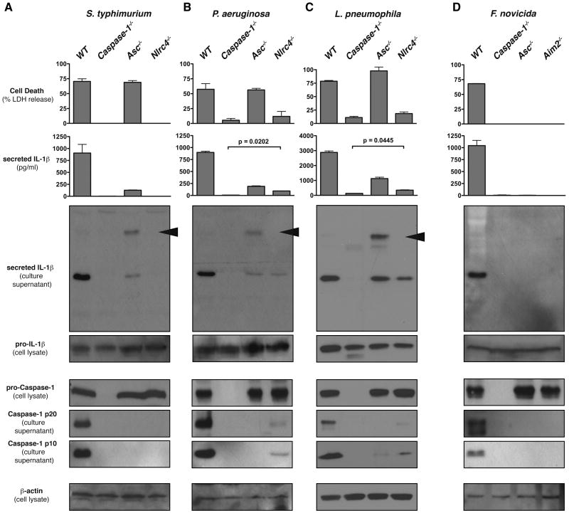
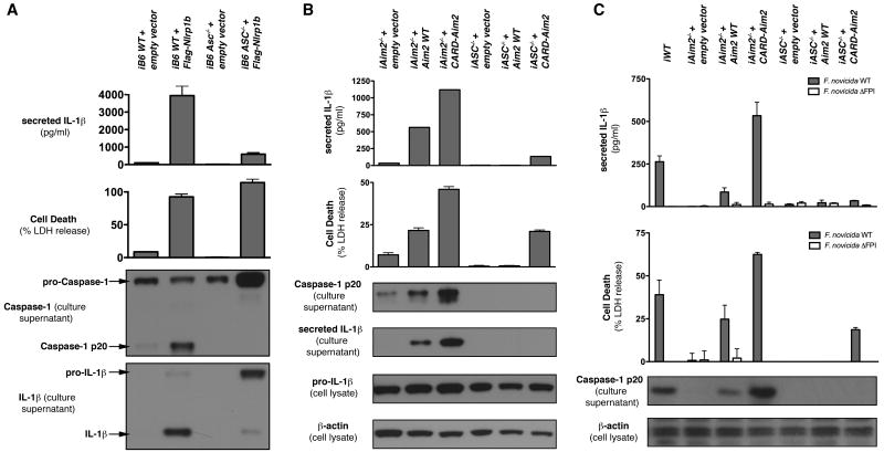
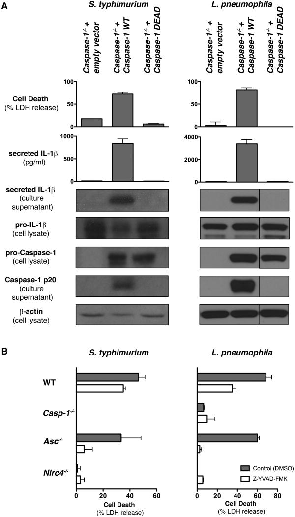
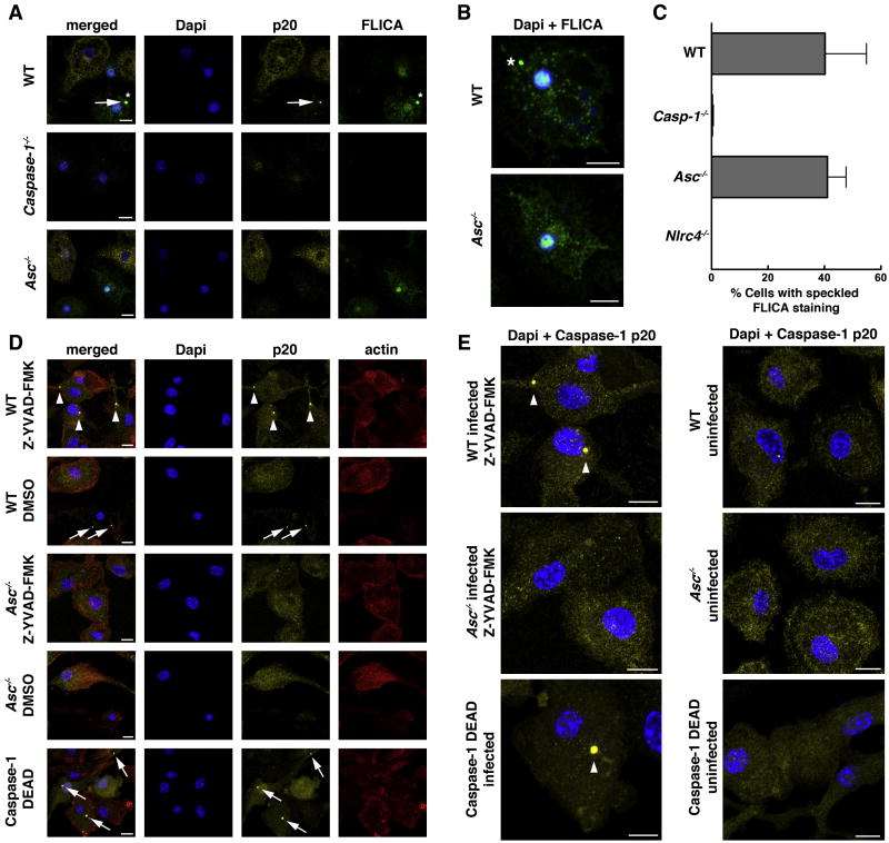
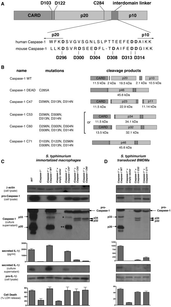
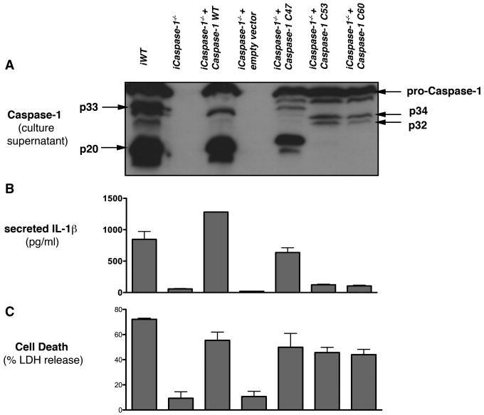
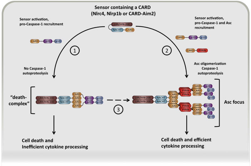
Activation of the cysteine protease Caspase-1 is a key event in the innate immune response to infections. Synthesized as a proprotein, Caspase-1 undergoes autoproteolysis within multiprotein complexes called inflammasomes. Activated Caspase-1 is required for proteolytic processing and for release of the cytokines interleukin-1β and interleukin-18, and it can also cause rapid macrophage cell death. We show that macrophage cell death and cytokine maturation in response to infection with diverse bacterial pathogens can be separated genetically and that two distinct inflammasome complexes mediate these events. Inflammasomes containing the signaling adaptor Asc form a single large "focus" in which Caspase-1 undergoes autoproteolysis and processes IL-1β/IL-18. In contrast, Asc-independent inflammasomes activate Caspase-1 without autoproteolysis and do not form any large structures in the cytosol. Caspase-1 mutants unable to undergo autoproteolysis promoted rapid cell death, but processed IL-1β/18 inefficiently. Our results suggest the formation of spatially and functionally distinct inflammasomes complexes in response to bacterial pathogens.


Figures

Figure 1
Figure 1. Nlrc4-dependent macrophage death occurs in the absence of Caspase-1 cleavage in Asc-deficient cells
Bone marrow-derived macrophages of the genotypes indicated were infected with (A) S. typhimurium for 2h at an MOI of 20, (B) P. aeruginosa PAO1 for 2h at an MOI of 10, (C) L. pneumophila for 4h at an MOI of 1 and (D) F. novicida for 6h at an MOI of 100. Cell death was determined by measuring LDH release. Secretion of mature IL-1β into the culture supernatant was determined by ELISA and western blotting. Release of processed Caspase-1 p10 and p20 subunits into the culture supernatant was determined by western blotting. Corresponding cell lysates were probed for pro-IL-1β, pro-Caspase-1 and β-actin. Graphs show the mean ± standard deviation (SD) of triplicate wells and are representative of at least three independent experiments. Arrowhead indicates pro-IL-1β in the supernatant.
Figure 2
Figure 2. CARD-containing sensors promote Asc-independent cell death
(A) Immortalized WT and Asc-/- macrophages derived from C57BL/6 mice expressing an empty vector control or the functional Nlrp1b allele from 129S1 mice were stimulated for 4h with Anthrax lethal toxin. (B, C) Immortalized WT, Aim2-/- or Asc-/- macrophages expressing an empty vector control, Aim2 WT or a CARD-Aim2 chimera were transfected with poly(dA:dT) (B) or infected with F. novicida WT or a mutant that does not activate the inflammasome (ΔFPI) (C). Cell death was determined by measuring LDH release. Secretion of IL-1β into the culture supernatant was determined by ELISA and western blotting. Release of processed Caspase-1 p20 into the culture supernatant was determined by western blotting. Corresponding cell lysates were probed for pro-IL-1β and β-actin. Graphs show the mean ± SD of triplicate wells and are representative of two independent experiments.
Figure 3
Figure 3. Catalytic activity of Caspase-1 is required for Asc-independent cell death
(A) Caspase-1-/- BMDMs expressing an empty vector control, WT Caspase-1 or the catalytically inactive mutation C284A (Caspase-1 DEAD) were infected with S. typhimurium for 2h at an MOI of 20 or L. pneumophila for 4h at an MOI of 1. Cell death was determined by measuring LDH release. Secretion of IL-1β into the culture supernatant was determined by ELISA and western blotting. Secretion of processed Caspase-1 p20 into the culture supernatant was determined by western blotting. Corresponding cell lysates were probed for pro-IL-1β, pro-Caspase-1 and β-actin. (B) BMDMs of the genotypes indicated were infected with S. typhimurium for 1h at an MOI of 20 or L. pneumophila for 2.5h at an MOI of 1 in the presence of Z-YVAD-FMK or DMSO (vehicle control). Graphs show the mean ± SD of triplicate wells and are representative of at least two independent experiments.
Figure 4
Figure 4. Differential subcellular localization of Caspase-1
(A) BMDMs of the genotypes indicated were infected with S. typhimurium for 2h at an MOI of 20, fixed and stained for DNA (Dapi), Caspase-1 p20 and with FLICA. Marked is an Asc focus stained by the Caspase-1 p20 antibody (arrow) or FLICA (asterisk). (B) Close up views of infected WT and Asc-/- macrophages stained for DNA (Dapi, blue) and with FLICA (green). (C) Percentage of cells with speckled, cytoplasmic FLICA staining. (D) WT and Asc-/- macrophages (treated with Z-YVAD-FMK or the vehicle control DMSO) or Caspase-1-/- macrophages expressing the catalytically inactive mutation C284A (Caspase-1 DEAD) were infected with S. typhimurium for 1h at an MOI of 20. Cells were fixed and stained for DNA (Dapi), Caspase-1 p20 and actin (Phalloidin). Indicated are Asc/Caspase-1 foci in the presence (arrowheads) or absence of the inhibitor (arrows). (E) Close up views of infected and uninfected WT and Asc-/- macrophages or Caspase-1-/- + Caspase-1 DEAD macrophages stained for DNA (Dapi, blue) and Caspase-1 p20 (yellow). Indicated are Asc/Caspase-1 foci (arrowheads). Cell counts in (C) were determined by counting twice 300 infected cells per sample. Images and cell counts are representative of at least two experiments. Scale bars in all images are 10 μm.
Figure 5
Figure 5. Caspase-1 autoproteolysis is necessary for efficient cytokine processing, but not for macrophage cell death in response to S. typhimurium infections
(A) Schematic representation of the domain organization of murine pro-Caspase-1, showing an alignment of the interdomain linker of human and murine Caspase-1. Putative cleavage sites and the active cysteine are indicated. (B) Schematic representation of different mutated Caspase-1 constructs, showing mutations and expected cleavage products. (C-D) Immortalized or bone-marrow derived Caspase-1-/- macrophages expressing the indicated Caspase-1 constructs were infected with S. typhimurium for 2h at an MOI of 20. Cell death was determined by measuring LDH release. Secretion of IL-1β into the culture supernatant was determined by ELISA and western blotting. Release of Caspase-1 into the culture supernatant was determined by western blotting. Corresponding cell lysates were probed for pro-Caspase-1, pro-IL-1β and β-actin. Graphs show the mean ± SD of triplicate wells and are representative of at least two independent experiments.
Figure 6
Figure 6. Caspase-1 processing is not required for macrophage death in response to F. novicida infections
Immortalized Caspase-1-/- macrophages expressing the indicated Caspase-1 constructs were infected with F. novicida for 9h at an MOI of 20. (A) Western blots for processed Caspase-1 p20 in culture supernatants. (B) ELISA for mature, secreted IL-1β in culture supernatants. (C) Cell death as determined by measuring LDH release. Arrows, arrowheads and asterisk indicate pro-Caspase-1 and different Caspase-1 auto-processing products. Results are representative of at least three independent experiments, error bars represent mean ± SD of triplicate wells.
Figure 7
Figure 7. Formation of spatially and functionally specialized inflammasome-complexes by CARD-containing receptors
1) Activation of CARD-containing sensors (Nlrc4, Nlrp1b or CARD-Aim2) by their respective stimuli induces dimerization of the sensor and recruitment of pro-Caspase-1 through CARD-CARD interactions. Proximity-driven dimerization in this “Death-complex” does not lead to autoproteolysis, however it activates pro-Caspase-1 sufficiently to promote cell death, but only inefficient cytokine processing. 2) Asc foci are formed when Asc is recruited in addition to pro-Caspase-1 to the activated sensor. Proximity driven dimerization of Asc promotes the recruitment of further Asc molecules by CARD-CARD and PYRIN-PYRIN interactions, leading to rapid oligomerization of Asc, thus forming the focus. In the Asc focus, conformational changes of the pro-Caspase-1 dimer allow for autoproteolytic processing into the p10 and p20 subunits. Only fully processed Caspase-1 is able to efficiently promote cytokine maturation. 3) Alternatively, the “Death-complexes” could serve as precursors or nuclei for Asc oligomerization and be absorbed by the Asc focus once it forms.

Similar articles

Cited by

References

    1. Amer A, Franchi L, Kanneganti TD, Body-Malapel M, Ozoren N, Brady G, Meshinchi S, Jagirdar R, Gewirtz A, Akira S, Nunez G. Regulation of Legionella phagosome maturation and infection through flagellin and host Ipaf. J Biol Chem. 2006;281:35217–35223. - PubMed
    1. Bao Q, Shi Y. Apoptosome: a platform for the activation of initiator caspases. Cell Death Differ. 2007;14:56–65. - PubMed
    1. Blasi E, Mathieson BJ, Varesio L, Cleveland JL, Borchert PA, Rapp UR. Selective immortalization of murine macrophages from fresh bone marrow by a raf/myc recombinant murine retrovirus. Nature. 1985;318:667–670. - PubMed
    1. Boatright KM, Renatus M, Scott FL, Sperandio S, Shin H, Pedersen IM, Ricci JE, Edris WA, Sutherlin DP, Green DR, Salvesen GS. A unified model for apical caspase activation. Mol Cell. 2003;11:529–541. - PubMed
    1. Boyden ED, Dietrich WF. Nalp1b controls mouse macrophage susceptibility to anthrax lethal toxin. Nat Genet. 2006;38:240–244. - PubMed

Publication types

MeSH terms