Comparative Study
. 2011 Apr;54(4):695-704.
doi: 10.1016/j.jhep.2010.10.004. Epub 2010 Oct 23.

Role of the EASL, RECIST, and WHO response guidelines alone or in combination for hepatocellular carcinoma: radiologic-pathologic correlation

Affiliations
Comparative Study

Role of the EASL, RECIST, and WHO response guidelines alone or in combination for hepatocellular carcinoma: radiologic-pathologic correlation

Ahsun Riaz et al. J Hepatol. 2011 Apr.

Abstract

Background & aims: We sought to study receiver-operating characteristics (ROC) of the European Association for the Study of the Liver (EASL), Response Evaluation Criteria in Solid Tumors (RECIST), and World Health Organization (WHO) guidelines for assessing response following locoregional therapies individually and in various combinations.

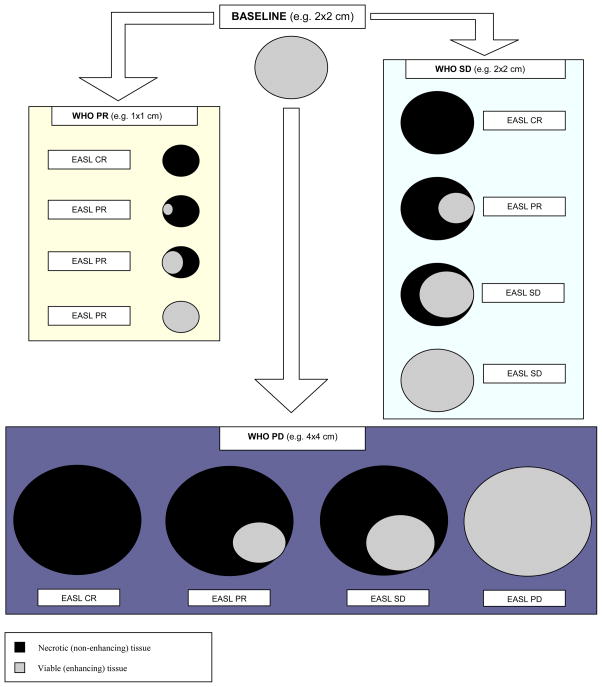
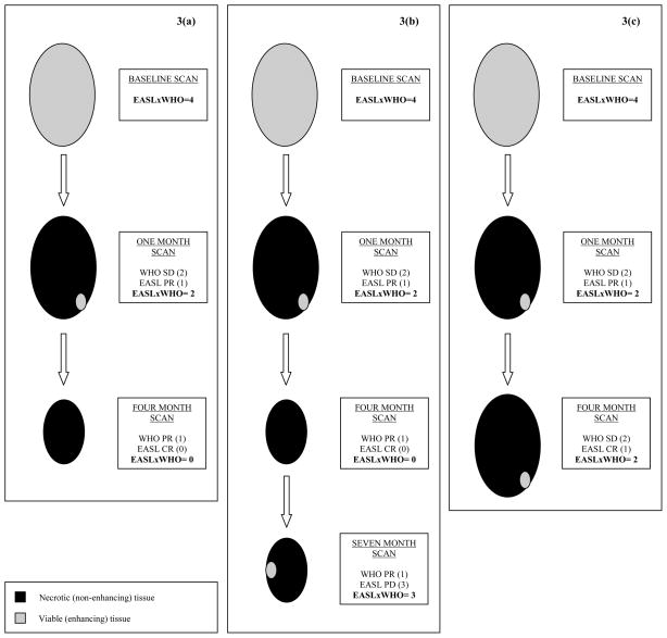
Methods: Eighty-one patients with hepatocellular carcinoma underwent liver explantation following locoregional therapies. Response was assessed using EASL, RECIST, and WHO. Kappa statistics were used to determine inter-method agreement. Uni/multivariate logistic regression analyses were performed to determine the variables predicting complete pathologic necrosis. Numerical values were assigned to the response classes: complete response=0, partial response=1, stable disease=2, and progressive disease=3. Various mathematical combinations of EASL and WHO were tested to calculate scores and their ROCs were studied using pathological examination of the explant as the gold standard.

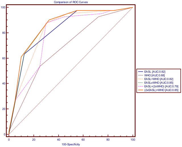
Results: Median times (95% CI) to the WHO, RECIST, and EASL responses were 5.3 (4-11.5), 5.6 (4-11.5), and 1.3months (1.2-1.5), respectively. Kappa coefficients for WHO/RECIST, WHO/EASL, and RECIST/EASL were 0.78, 0.28, and 0.31, respectively. EASL response demonstrated significant odds ratios for predicting complete pathologic necrosis on uni/multivariate analyses. Calculated areas under the ROC curves were: RECIST: 0.63, WHO: 0.68, EASL: 0.82, EASL+WHO: 0.82, EASL×WHO: 0.85, EASL+(2×WHO): 0.79 and (2×EASL)+WHO: 0.85. An EASL×WHO Score of ⩽1 had 90.2% sensitivity for predicting complete pathologic necrosis.

Conclusions: The product of WHO and EASL demonstrated better ROC than the individual guidelines for assessment of tumor response. EASL×WHO scoring system provides a simple and clinically applicable method of response assessment following locoregional therapies for hepatocellular carcinoma.


Conflict of interest statement

Conflict of Interest: No conflicts of interest.

Figures

Figure 1
Figure 1
Schematic representation of the World Health Organization (WHO) and European Association for Study of the Liver (EASL) guidelines. The WHO guidelines suggest measurement of the whole tumor irrespective of the amount of necrosis (black) seen. The EASL guidelines suggest measurement of the enhancing tissue only (grey). This figure represents the discordance between the two guidelines.
Figure 2
Figure 2
Figure demonstrating comparison of receiver-operating characteristics (ROC) curves using the various scoring systems. The following scoring systems were analyzed: Response Evaluation Criteria for Solid Tumors (RECIST); World Health Organization (WHO) guidelines; European Association for Study of the Liver (EASL); EASL + WHO; EASL × WHO; EASL + (2xWHO); (2xEASL) + WHO.
Figure 3
Figure 3
Examples using EASLxWHO scoring systems. A patient is diagnosed with a 2x2 cm2 HCC. The baseline EASLxWHO Score assigned to this lesion would be 4 [SD (2) by both guidelines; 2x2=4]. 3(a): The one-month post-treatment scan shows that the size of the lesion remains the same but the lesion shows EASL PR (EASLxWHO Score=1x2=2). This decrease in the EASLxWHO Score at the one-month scan would demonstrate response in the treated lesion. Clinically, this would translate into observation of the patient with routine scheduled follow-up. The four-month scan shows that the size of the lesion is now 1x1 cm2 and the lesion now shows an EASL CR (EASLxWHO Score=0x1=0). In this patient the EASLxWHO Scores allocated to this patient were 4 → 2 → 0 at baseline, one-month and four-month follow-up scans, respectively; the odds of this lesion having complete pathologic necrosis are high. This decrease in the EASLxWHO Score would again translate into observation of the patient and routine follow-up. 3(b): In the same patient as the one represented in Figure 3a, the seven-month scan shows that the lesion is still 1x1 cm2 but now shows reappearance of enhancing tissue which would be classified as EASL PD (EASLxWHO Score=1x3=3). In this patient with an initial response, there is subsequent progression as seen in the EASLxWHO Scores of 4 → 2 → 0 → 3 at baseline, one-month, four-month and seven-month follow-up scans, respectively. Now that the score is >1, this would signify progression and may translate into clinical interventions such as re-treatment. 3(c): Another clinical scenario may be presented as follows using the same patient example as above with similar findings on the one-month post-treatment scan (EASLxWHO Score=1x2=2). However, the findings on the four-month post-treatment scan show that the lesion remained 2x2 cm2 and there is still enhancing tissue in the lesion (EASL PR), the EASLxWHO Score remains 2. In this patient the allocated EASLxWHO Scores were 4 → 2 → 2 at baseline, one-month and four-month follow-up scans, respectively. This stability in the score between the one-month and the four-month scan may demonstrate treatment failure due to incomplete targeting of the tumor.
Figure 4
Figure 4
4(a) Contrast-enhanced MRI demonstrates a large right lobe biopsy-proven HCC. Although the baseline scan clearly demonstrates a viable tumor, there is a moderate amount of necrosis at baseline. Quantifying the amount of enhancing tissue (viable hepatocellular carcinoma) at baseline without using computer software applying EASL guidelines would be difficult given the irregular and ill-defined interface between viable disease and necrotic tissue. 4(b) Post-treatment MRI demonstrates a decrease in enhancing tissue representing a clear EASL response by subjective assessment; however the exact quantification of necrosis versus viable tissue is again difficult because of the persistent irregularity in pattern of contrast enhancement.
Figure 4
Figure 4
4(a) Contrast-enhanced MRI demonstrates a large right lobe biopsy-proven HCC. Although the baseline scan clearly demonstrates a viable tumor, there is a moderate amount of necrosis at baseline. Quantifying the amount of enhancing tissue (viable hepatocellular carcinoma) at baseline without using computer software applying EASL guidelines would be difficult given the irregular and ill-defined interface between viable disease and necrotic tissue. 4(b) Post-treatment MRI demonstrates a decrease in enhancing tissue representing a clear EASL response by subjective assessment; however the exact quantification of necrosis versus viable tissue is again difficult because of the persistent irregularity in pattern of contrast enhancement.
Figure 5
Figure 5
5(a) This is an enhancing hepatoma in segment 5/8. 5(b) Following treatment, one can clearly see the radiation effect along the vascular planes of segment 5/8. Because of the slight irregular mural enhancement, this was labeled as EASL PR, illustrating the conservative methodology used throughout the analysis.
Figure 5
Figure 5
5(a) This is an enhancing hepatoma in segment 5/8. 5(b) Following treatment, one can clearly see the radiation effect along the vascular planes of segment 5/8. Because of the slight irregular mural enhancement, this was labeled as EASL PR, illustrating the conservative methodology used throughout the analysis.

Similar articles

Cited by

  • The physics of radioembolization.
    Bastiaannet R, Kappadath SC, Kunnen B, Braat AJAT, Lam MGEH, de Jong HWAM. Bastiaannet R, et al. EJNMMI Phys. 2018 Nov 2;5(1):22. doi: 10.1186/s40658-018-0221-z. EJNMMI Phys. 2018. PMID: 30386924 Free PMC article. Review.
  • Clinical significance of AFP and PIVKA-II responses for monitoring treatment outcomes and predicting prognosis in patients with hepatocellular carcinoma.
    Park H, Park JY. Park H, et al. Biomed Res Int. 2013;2013:310427. doi: 10.1155/2013/310427. Epub 2013 Dec 29. Biomed Res Int. 2013. PMID: 24455683 Free PMC article.
  • Squaring the circle of selection and allocation in liver transplantation for HCC: An adaptive approach.
    Mazzaferro V. Mazzaferro V. Hepatology. 2016 May;63(5):1707-17. doi: 10.1002/hep.28420. Epub 2016 Feb 26. Hepatology. 2016. PMID: 26703761 Free PMC article. No abstract available.
  • Radioembolization for primary and metastatic liver cancer.
    Memon K, Lewandowski RJ, Kulik L, Riaz A, Mulcahy MF, Salem R. Memon K, et al. Semin Radiat Oncol. 2011 Oct;21(4):294-302. doi: 10.1016/j.semradonc.2011.05.004. Semin Radiat Oncol. 2011. PMID: 21939859 Free PMC article. Review.
  • [Diagnosis of and therapy for hepatocellular carcinoma].
    Greten TF, Malek NP, Schmidt S, Arends J, Bartenstein P, Bechstein W, Bernatik T, Bitzer M, Chavan A, Dollinger M, Domagk D, Drognitz O, Düx M, Farkas S, Folprecht G, Galle P, Geißler M, Gerken G, Habermehl D, Helmberger T, Herfarth K, Hoffmann RT, Holtmann M, Huppert P, Jakobs T, Keller M, Klempnauer J, Kolligs F, Körber J, Lang H, Lehner F, Lordick F, Lubienski A, Manns MP, Mahnken A, Möhler M, Mönch C, Neuhaus P, Niederau C, Ocker M, Otto G, Pereira P, Pott G, Riemer J, Ringe K, Ritterbusch U, Rummeny E, Schirmacher P, Schlitt HJ, Schlottmann K, Schmitz V, Schuler A, Schulze-Bergkamen H, von Schweinitz D, Seehofer D, Sitter H, Straßburg CP, Stroszczynski C, Strobel D, Tannapfel A, Trojan J, van Thiel I, Vogel A, Wacker F, Wedemeyer H, Wege H, Weinmann A, Wittekind C, Wörmann B, Zech CJ. Greten TF, et al. Z Gastroenterol. 2013 Nov;51(11):1269-326. doi: 10.1055/s-0033-1355841. Epub 2013 Nov 15. Z Gastroenterol. 2013. PMID: 24243572 Free PMC article.

References

    1. El-Serag HB. Hepatocellular carcinoma and hepatitis C in the United States. Hepatology. 2002;36(5 Suppl 1):S74–83. - PubMed
    1. Mazzaferro V, Regalia E, Doci R, Andreola S, Pulvirenti A, Bozzetti F, et al. Liver transplantation for the treatment of small hepatocellular carcinomas in patients with cirrhosis. N Engl J Med. 1996;334(11):693–699. - PubMed
    1. Llovet JM, Ricci S, Mazzaferro V, Hilgard P, Gane E, Blanc JF, et al. Sorafenib in advanced hepatocellular carcinoma. N Engl J Med. 2008;359(4):378–390. - PubMed
    1. Cheng AL, Kang YK, Chen Z, Tsao CJ, Qin S, Kim JS, et al. Efficacy and safety of sorafenib in patients in the Asia-Pacific region with advanced hepatocellular carcinoma: a phase III randomised, double-blind, placebo-controlled trial. Lancet Oncol. 2009;10(1):25–34. - PubMed
    1. Salem R, Lewandowski RJ, Mulcahy MF, Riaz A, Ryu RK, Ibrahim S, et al. Radioembolization for hepatocellular carcinoma using Yttrium-90 microspheres: a comprehensive report of long-term outcomes. Gastroenterology. 2010;138(1):52–64. - PubMed

Publication types