

A novel role for progesterone and progesterone receptor membrane component 1 in regulating spindle microtubule stability during rat and human ovarian cell mitosis
- PMID: 21148105
- PMCID: PMC3062038
- DOI: 10.1095/biolreprod.110.088385
A novel role for progesterone and progesterone receptor membrane component 1 in regulating spindle microtubule stability during rat and human ovarian cell mitosis
Abstract
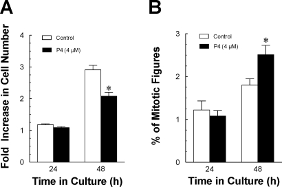
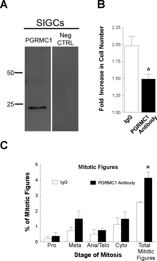
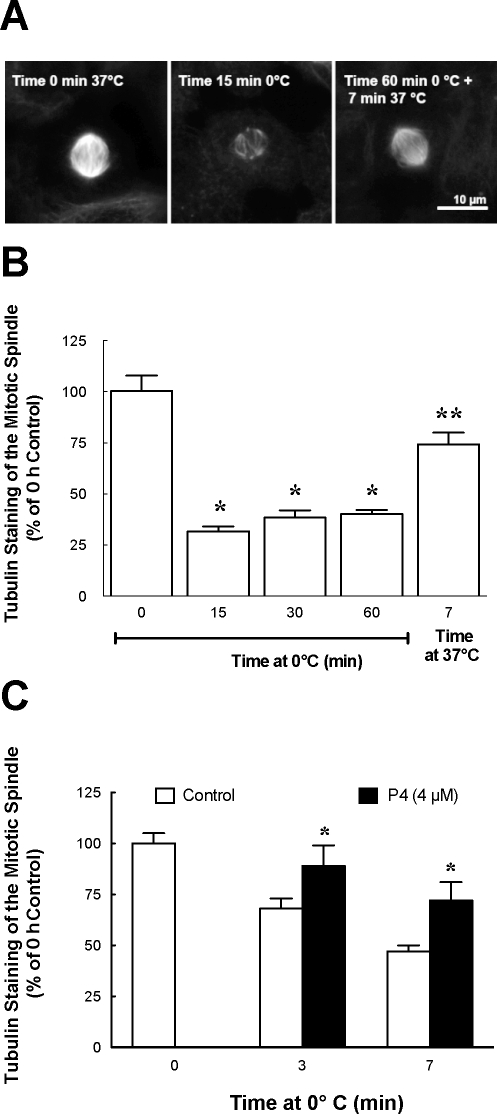
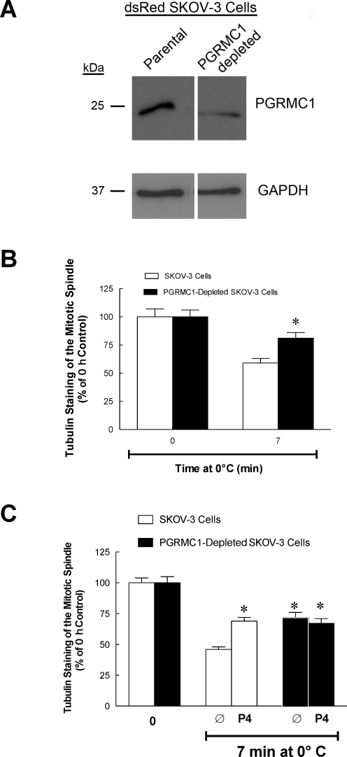
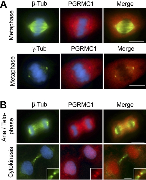
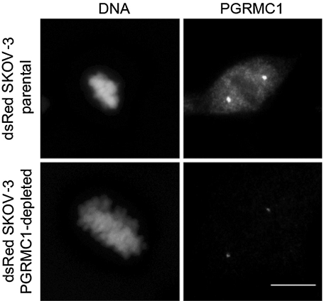
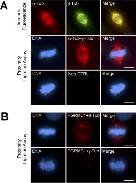
The present studies were designed to assess the roles of progesterone (P4) and Progesterone Receptor Membrane Component 1 (PGRMC1) in regulating mitosis of spontaneously immortalized granulosa cells (SIGCs) and ovarian cancer cells, SKOV-3 cells. Because PGRMC1 has been detected among the proteins of the human mitotic spindle, we theorized that P4 and PGRMC1 could affect mitosis through a microtubule-dependent process. The present study confirms that SIGC growth is slowed by either P4 treatment or transfection of a PGRMC1 antibody. In both cases, slower cell proliferation was accompanied by an increased percentage of mitotic cells, which is consistent with a P4-induced prolongation of the M phase of the cell cycle. In addition, P4 increased the stability of the spindle microtubules, as assessed by the rate of beta-tubulin disassembly in response to cooling. Also, P4 increased spindle microtubule stability of SKOV-3 cells. This effect was mimicked by the depletion of PGRMC1 in these cells. Importantly, P4 did not increase the stability of the microtubules over that observed in PGRMC1-depleted SKOV-3 cells. Immunofluorescent analysis revealed that PGRMC1 is distributed to the spindle apparatus as well as to the centrosomes at metaphase. Further in situ proximity ligation assay revealed that PGRMC1 interacted with beta-tubulin. Taken together, these results suggest that P4 inhibits mitosis of ovarian cells by increasing the stability of the mitotic spindle. Moreover, P4's actions appear to be dependent on PGRMC1's function within the mitotic spindle.
Figures
Similar articles
-
Progesterone membrane receptor component 1 expression in the immature rat ovary and its role in mediating progesterone's antiapoptotic action.Endocrinology. 2006 Jun;147(6):3133-40. doi: 10.1210/en.2006-0114. Epub 2006 Mar 2. Endocrinology. 2006. PMID: 16513825
-
Progesterone receptor membrane component-1 (PGRMC1) and PGRMC-2 interact to suppress entry into the cell cycle in spontaneously immortalized rat granulosa cells.Biol Reprod. 2014 Nov;91(5):104. doi: 10.1095/biolreprod.114.122986. Epub 2014 Sep 24. Biol Reprod. 2014. PMID: 25253729 Free PMC article.
-
Plasminogen activator inhibitor 1 RNA-binding protein interacts with progesterone receptor membrane component 1 to regulate progesterone's ability to maintain the viability of spontaneously immortalized granulosa cells and rat granulosa cells.Biol Reprod. 2013 Jan 25;88(1):20. doi: 10.1095/biolreprod.112.103036. Print 2013 Jan. Biol Reprod. 2013. PMID: 23242527 Free PMC article.
-
Non-canonical progesterone signaling in granulosa cell function.Reproduction. 2014 Apr 8;147(5):R169-78. doi: 10.1530/REP-13-0582. Print 2014 May. Reproduction. 2014. PMID: 24516175 Free PMC article. Review.
-
Progesterone signaling mediated through progesterone receptor membrane component-1 in ovarian cells with special emphasis on ovarian cancer.Steroids. 2011 Aug;76(9):903-9. doi: 10.1016/j.steroids.2011.02.011. Epub 2011 Mar 1. Steroids. 2011. PMID: 21371489 Free PMC article. Review.
Cited by
-
Searching for the 'X' factor: investigating the genetics of primary ovarian insufficiency.J Ovarian Res. 2024 Nov 28;17(1):238. doi: 10.1186/s13048-024-01555-5. J Ovarian Res. 2024. PMID: 39609914 Free PMC article. Review.
-
PGRMC1-dependent lipophagy promotes ferroptosis in paclitaxel-tolerant persister cancer cells.J Exp Clin Cancer Res. 2021 Nov 8;40(1):350. doi: 10.1186/s13046-021-02168-2. J Exp Clin Cancer Res. 2021. PMID: 34749765 Free PMC article.
-
Evidence for a genomic mechanism of action for progesterone receptor membrane component-1.Steroids. 2012 Aug;77(10):1007-12. doi: 10.1016/j.steroids.2012.01.013. Epub 2012 Feb 1. Steroids. 2012. PMID: 22326699 Free PMC article. Review.
-
Insights on the Role of PGRMC1 in Mitotic and Meiotic Cell Division.Cancers (Basel). 2022 Nov 23;14(23):5755. doi: 10.3390/cancers14235755. Cancers (Basel). 2022. PMID: 36497237 Free PMC article. Review.
-
PGRMC1 and PGRMC2 in uterine physiology and disease.Front Neurosci. 2013 Sep 19;7:168. doi: 10.3389/fnins.2013.00168. eCollection 2013. Front Neurosci. 2013. PMID: 24065879 Free PMC article.
References
-
- Mahesh VB, Muldoon TG. Integration of the effects of estradiol and progesterone in the modulation of gonadotropin secretion. J Steroid Biochem 1987; 27: 665 675 - PubMed
-
- Muldoon TG, Mahesh VB. Receptor-weighted mechanistic approach to analysis of the actions of estrogen and progesterone on gonadotropin secretion. Adv Exp Med Biol 1987; 219: 47 64 - PubMed
-
- Kurita T, Lee K, Saunders PT, Cooke PS, Taylor JA, Lubahn DB, Zhao C, Makela S, Gustafsson JA, Dahiya R, Cunha GR. Regulation of progesterone receptors and decidualization in uterine stroma of the estrogen receptor-alpha knockout mouse. Biol Reprod 2001; 64: 272 283 - PubMed
-
- Spencer TE, Johnson GA, Burghardt RC, Bazer FW. Progesterone and placental hormone actions on the uterus: insights from domestic animals. Biol Reprod 2004; 71: 2 10 - PubMed
-
- Peluso JJ. Multiplicity of progesterone's actions and receptors in the mammalian ovary. Biol Reprod 2006; 75: 2 8 - PubMed
Publication types
MeSH terms
Substances
Grants and funding
LinkOut - more resources
Full Text Sources
Other Literature Sources
Research Materials
