. 2011 Mar;25(3):1028-39.
doi: 10.1096/fj.10-168799. Epub 2010 Dec 8.

Novel insights into the regulation of skeletal muscle protein synthesis as revealed by a new nonradioactive in vivo technique

Affiliations

Novel insights into the regulation of skeletal muscle protein synthesis as revealed by a new nonradioactive in vivo technique

Craig A Goodman et al. FASEB J. 2011 Mar.

Abstract

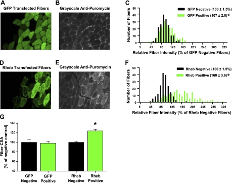
In this study, the principles of surface sensing of translation (SUnSET) were used to develop a nonradioactive method for ex vivo and in vivo measurements of protein synthesis (PS). Compared with controls, we first demonstrate excellent agreement between SUnSET and a [(3)H]phenylalanine method when detecting synergist ablation-induced increases in skeletal muscle PS ex vivo. We then show that SUnSET can detect the same synergist ablation-induced increase in PS when used in vivo (IV-SUnSET). In addition, IV-SUnSET detected food deprivation-induced decreases in PS in the heart, kidney, and skeletal muscles, with similar changes being visualized with an immunohistochemical version of IV-SUnSET (IV-IHC-SUnSET). By combining IV-IHC-SUnSET with in vivo transfection, we demonstrate that constitutively active PKB induces a robust increase in skeletal muscle PS. Furthermore, transfection with Ras homolog enriched in brain (Rheb) revealed that a PKB-independent activation of mammalian target of rapamycin is also sufficient to induce an increase in skeletal muscle PS. Finally, IV-IHC-SUnSET exposed the existence of fiber type-dependent differences in skeletal muscle PS, with PS in type 2B and 2X fibers being significantly lower than that in type 2A fibers within the same muscle. Thus, our nonradioactive method allowed us to accurately visualize and quantify PS under various ex vivo and in vivo conditions and revealed novel insights into the regulation of PS in skeletal muscle.


Figures

Figure 1.
Figure 1.
SA induces hypertrophy, increased rRNA, and increased protein synthesis as measured ex vivo with radioactive- and SUnSET-based techniques. PLT muscles were subjected to sham (control) or SA surgical procedures and after 7 d were assessed for changes in parameters. A) Muscle weight (MW)/body weight (BW) ratio (n=5/group). B) Total protein content (n=5/group). C) Total RNA/MW (n=5/group). D) Relative 28S and 18S rRNA/MW (n=5/group). E–G) Ex vivo rates of protein synthesis (n=3/group). E, F) SUnSET measurements of protein synthesis were performed by incubating muscles in an organ culture bath with medium containing puromycin as described in Materials and Methods. E) Representative image of WB analysis for puromycin followed by Coomassie Blue staining to verify equal loading of proteins. Note that the specificity of the anti-puromycin blot was demonstrated by a sample that was not incubated with puromycin (Puro, far left lane). F) Quantification of the puromycin-labeled peptides, expressed as a percentage of the values obtained in the control group. RLU, relative light units. G) Radioactive measurements of protein synthesis rate were performed by incubating muscles in an organ culture bath with medium containing a flooding dose of [3H]phenylalanine as described in Materials and Methods. All values are means + se. *P < 0.05 vs. control.
Figure 2.
Figure 2.
SUnSET measurements of enhanced protein synthesis in the absence of changes in rRNA. Serum-starved C2C12 myoblasts were stimulated with or without 100 nM insulin (INS) for 90 min, and rates of protein synthesis were measured with the SUnSET technique described in Materials and Methods. A) Representative image of WB analysis for puromycin followed by Coomassie Blue staining to verify equal loading of proteins. B) Quantification of the puromycin-labeled peptides (n=8 wells/group from 3 independent experiments). RLU, relative light units. C) Relative concentration of 28S and 18S rRNA per cell culture well (n=6 wells/group from 3 independent experiments). All values are expressed as a percentage of the values obtained in control samples and are presented as means + se. *P < 0.05 vs. control.
Figure 3.
Figure 3.
SA induces protein synthesis in vivo as measured with IV-SUnSET. PLT muscles were subjected to sham (control) or SA surgical procedures, and after 7 d in vivo rates of protein synthesis were determined with the IV-SUnSET technique described in Materials and Methods. A) Representative image of WB analysis for puromycin-labeled peptides followed by Coomassie Blue staining to verify equal loading of proteins. B) Quantification of the puromycin-labeled peptides, expressed as a percentage of the values obtained in the control group (n=3/group). RLU, relative light units. C) Measurement of the free puromycin concentration (n=6/group). Values are means + se. *P < 0.05 vs. control.
Figure 4.
Figure 4.
Food deprivation induces a decrease in protein synthesis as determined with IV-SUnSET and IV-IHC-SUnSET. Mice were fed ad libitum (control) or deprived of food for 48 h (fasted), and the in vivo rates of protein synthesis in the heart (A, B), kidney (C, D), and TA skeletal muscle (E, F) were measured with IV-SUnSET as described in Materials and Methods. A, C, E) Representative images of WB analysis for puromycin (Puro) followed by Coomassie Blue (CB) staining to verify equal loading of proteins. B, D, F) Quantification of the puromycin-labeled peptides, expressed as a percentage of the values obtained in the control group. RLU, relative light units. A–F) All values are means ± se (for heart, n=8/group; for kidney and TA, n=4–5/group). G, H) In vivo rates of protein synthesis in the TA muscle were determined with an IV-IHC-SUnSET as described in Materials and Methods. G) Representative image of cross sections from TA muscles that were subjected to control or food-deprivation conditions, mounted adjacent to one another on a slide, and then subjected to IHC for puromycin. H) Puromycin staining intensity in individual fibers from both control and food-deprivation sections was expressed relative to the mean puromycin signal intensity of fibers in control sections and then plotted on a histogram. Average ± se staining intensity is indicated next to group labels in legend; n = 300 fibers/group from 5 independent pairs of muscles. *P < 0.05 vs. control.
Figure 5.
Figure 5.
In vivo overexpression of CA-PKB induces an increase in protein synthesis as determined with IV-IHC-SUnSET. TA muscles were transfected with plasmid DNA encoding the GFP (A–C) or HA-tagged CA-PKB (D–F). At 3 d after transfection, muscles were subjected to IV-IHC-SUnSET by staining for puromycin and GFP or the HA tag, respectively. A) Representative image of the signal for GFP positive fibers. B) Grayscale image of the signal for puromycin in the same section shown in A. D) Representative image of the signal for CA-PKB positive fibers. E) Grayscale image of the signal for puromycin in the same section shown in D. C, F) Puromycin staining intensity in both transfected (positive) and nontransfected (negative) fibers, expressed relative to the mean value obtained in nontransfected (negative) fibers from the same section; values are plotted on histograms for GFP-transfected muscles (C) and CA-PKB-transfected muscles (F). Values next to group labels in legend are means ± se; n = 180 transfected and nontransfected fibers/group from 3 independent muscles. *P < 0.05 vs. control.
Figure 6.
Figure 6.
IV-IHC-SUnSET reveals that PKB-independent activation of mTOR is sufficient to induce an increase in skeletal muscle protein synthesis and hypertrophy. TA muscles were transfected with plasmid DNA encoding the GFP (A–C) or HA-tagged Rheb (D–F). At 4 d after transfection, muscles were subjected to IV-IHC-SUnSET by staining for puromycin and GFP or the HA tag, respectively. A) Representative image of the signal for GFP-positive fibers. B) Grayscale image of the signal for puromycin in the same section shown in A. D) Representative image of signal for Rheb-positive fibers. E) Grayscale image of the signal for puromycin in the same section shown in D. C, F) Puromycin staining intensity in both transfected (positive) and nontransfected (negative) fibers, expressed relative to the mean value obtained in nontransfected (negative) fibers from the same section; values are plotted on histograms for GFP-transfected muscles (C) and Rheb-transfected muscles (F). G) Cross-sectional area (CSA) of the transfected fibers was measured and expressed relative to the CSA of nontransfected fibers in the same section. All values are means ± se; n = 180 transfected and nontransfected fibers/group from 3 independent muscles. *P < 0.05 vs. nontransfected fibers..
Figure 7.
Figure 7.
IV-IHC-SUnSET reveals skeletal muscle fiber type-dependent differences in protein synthesis. A, D, G) PLT muscles were subjected to IV-IHC-SUnSET by triple staining for puromycin (Puro, red), type 2A fibers (green), and either type 2B (A), type 2X (D), or type 1 fibers (blue; G). B, E, H) Grayscale image of the puromycin signal in the same section as shown in A, D, and G, respectively. C, F, I) Puromycin staining intensity in type 2A fibers and either type 2B, type 2X, or type 1 fibers, expressed relative to the mean value obtained in type 2A fibers from a given section; values are plotted on histograms for type 2B (C), type 2X (F), and type 1 fibers (I). Values are means ± se; n = 87–120 fibers/group from 4 independent muscles. *P < 0.05 vs. type 2A fibers.

References

    1. Wolfe R. R., Chinkes D. L. (2005) Isotope Tracers in Metabolic Research, John Wiley & Sons, Hoboken, NJ, USA
    1. Schmidt E. K., Clavarino G., Ceppi M., Pierre P. (2009) SUnSET, a nonradioactive method to monitor protein synthesis. Nat. Methods 6, 275–277 - PubMed
    1. Nathans D. (1964) Puromycin inhibition of protein synthesis: incorporation of puromycin into peptide chains. Proc. Natl. Acad. Sci. U. S. A. 51, 585–592 - PMC - PubMed
    1. Nakano K., Hara H. (1979) Measurement of the protein-synthetic activity in vivo of various tissues in rats by using [3H]puromycin. Biochem. J. 184, 663–668 - PMC - PubMed
    1. Sandri M. (2008) Signaling in muscle atrophy and hypertrophy. Physiology (Bethesda) 23, 160–170 - PubMed

Publication types

MeSH terms