. 2011 Feb 18;286(7):5126-35.
doi: 10.1074/jbc.M110.180711. Epub 2010 Dec 8.

Interactions of perilipin-5 (Plin5) with adipose triglyceride lipase

Affiliations

Interactions of perilipin-5 (Plin5) with adipose triglyceride lipase

James G Granneman et al. J Biol Chem. .

Abstract

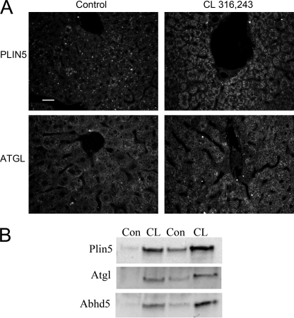
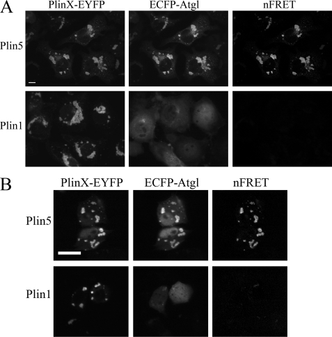
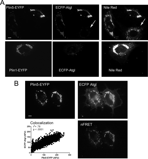
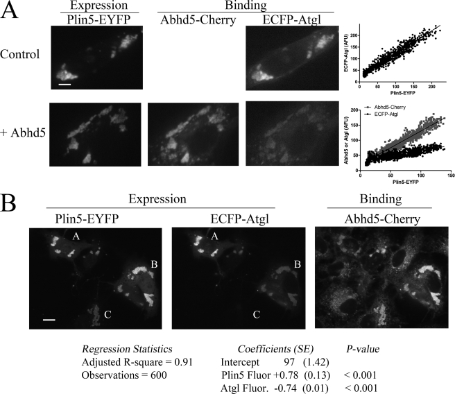
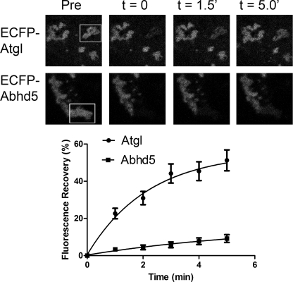
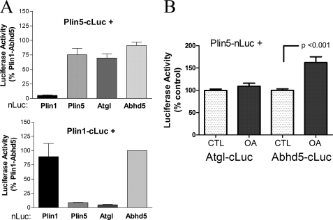
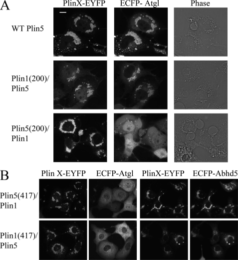
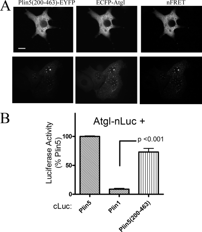
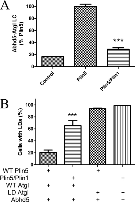
Members of the perilipin family of lipid droplet scaffold proteins are thought to play important roles in tissue-specific regulation of triglyceride metabolism, but the mechanisms involved are not fully understood. Present results indicate that adipose triglyceride lipase (Atgl) interacts with perilipin-5 (Plin5) but not perilipin-1 (Plin1). Protein interaction assays in live cells and in situ binding experiments showed that Atgl and its protein activator, α-β-hydrolase domain-containing 5 (Abhd5), each bind Plin5. Surprisingly, competition experiments indicated that individual Plin5 molecules bind Atgl or Abhd5 but not both simultaneously. Thus, the ability of Plin5 to concentrate these proteins at droplet surfaces involves binding to different Plin5 molecules, possibly in an oligomeric complex. The association of Plin5-Abhd5 complexes on lipid droplet surfaces was more stable than Plin5-Atgl complexes, and oleic acid treatment selectively promoted the interaction of Plin5 and Abhd5. Analysis of chimeric and mutant perilipin proteins demonstrated that amino acids 200-463 are necessary and sufficient to bind both Atgl and Abhd5 and that the C-terminal 64 amino acids of Plin5 are critical for the differential binding of Atgl to Plin5 and Plin1. Mutant Plin5 that binds Abhd5 but not Atgl was defective in preventing neutral lipid accumulation compared with wild type Plin5, indicating that the ability of Plin5 to concentrate these proteins on lipid droplets is critical to functional Atgl activity in cells.


Figures

FIGURE 1.
FIGURE 1.
Colocalization of Plin5, Atgl, and Abhd5 on lipid droplets of steatotic liver. A, immunofluorescence localization of Plin5 and Atgl was performed on liver sections of control mice and mice treated with CL to produce hepatic steatosis. B, immunoblot detection of Plin5, Atgl, and Abhd5 in lipid droplet fractions of control (Con) and CL-treated mice. Bar, 20 μm.
FIGURE 2.
FIGURE 2.
ECFP-Atgl colocalizes and interacts with Plin5 but not Plin1. A, interaction of ECFP-Atgl and Plin5-EYFP in COS7 cells. COS7 cells were transiently transfected with ECFP-Atgl and EYFP-tagged Plin5 or Plin1 and imaged by confocal microscopy. ECFP-Atgl was highly targeted to lipid droplets containing Plin5-EYFP but not to droplets containing Plin1. The colocalization of ECFP-Atgl and Plin5-EYFP supported FRET on lipid droplets. Bar, 10 μm. B, interaction of ECFP-Atgl and Plin5-EYFP in H4IIe hepatoma cells. Cotransfection of Plin5-EYFP and ECFP-Atgl leads to colocalization on lipid droplets and FRET between fluorescent proteins. In contrast, Plin1-EYFP did not support droplet targeting of ECFP-Atgl or FRET. Bar, 10 μm.
FIGURE 3.
FIGURE 3.
ECFP-Atgl selectively binds Plin5-EYFP in permeabilized cells. A, binding of recombinant ECFP-Atgl in permeabilized COS7 cells expressing EYFP-tagged Plin5 or Plin1. Permeabilized cells were incubated with the same preparation of ECFP-Atgl, and all capture parameters were the same for each fluorescence channel. ECFP-Atgl bound to lipid droplets (post-stained with Nile Red) of cells transfected with Plin5, but not to untransfected cells (arrow) or cells expressing Plin1. B, cells expressing Plin5-EYFP were permeabilized and bound with recombinant ECFP-Atgl as detailed under “Experimental Procedures.” Atgl bound in direct proportion to Plin5 concentration as demonstrated by linear regression of pixel intensities (see also A). In addition, bound ECFP-Atgl supported FRET with droplet-associated Plin5, indicating close interaction. Bar, 10 μm.
FIGURE 4.
FIGURE 4.
Mutually exclusive binding of Atgl and Abdh5 to Plin5. A, binding of Abhd5-Cherry suppresses binding of ECFP-Atgl to Plin5-EYFP. COS7 cells expressing Plin5-EYFP were fixed and permeabilized and then bound with ECFP-Atgl alone (top panel) or with Abhd5-Cherry (bottom panel). Images are from a representative experiment, and all capture parameters were equal for each fluorescence channel. Coincubation with Abhd5-Cherry significantly reduced binding of ECFP-Atgl as assessed by the regression analysis (p < 0.001). B, binding of ECFP-Atgl to Plin5-EYFP prevents binding of Abhd5-Cherry. COS7 cells coexpressing variable amounts of ECFP-Atgl and Plin5-EYFP were fixed and permeabilized and then bound with Abhd5-Cherry. A, B, and C are cells with variable amounts of bound ECFP-Atgl. Binding was assessed by the intensity of bound Abhd5-Cherry fluorescence as a function of Plin5 fluorescence (Plin5 Fluor.) and Atgl fluorescence (Atgl Fluor.) intensities. Binding was directly proportional to Plin 5 fluorescence (slope = +0.78) and inversely proportional to Atgl fluorescence (slope = −0.74). Bar, 10 μm.
FIGURE 5.
FIGURE 5.
Complexes of ECFP-Abhd5 and Plin5-EYFP on lipid droplets are more stable than complexes of ECFP-Atgl and Plin5-EYFP. Top panel, confocal micrographs of cells before (Pre) and after photobleaching of indicated region of interest (box). Bottom panel, summary of recovery in 10–14 bleaching experiments. Composite data were fit to a single exponential association function by nonlinear regression.
FIGURE 6.
FIGURE 6.
Protein-protein interaction of Atgl with Plin5 and Plin1 assessed by luciferase complementation assays. A, Plin5 interacts with Atgl, but Plin1 does not. 293T cells were transiently transfected with Plin5 or Plin1 tagged with the C-terminal fragment of Gaussia princeps luciferase (cLuc) and the complementary N-terminal luciferase (nLuc) fragment fused to potential binding partners. Complementation of luciferase activity was used to evaluate protein-protein interaction. Plin5 interacted with Atgl, Abhd5, and itself but not with Plin1. Plin1 interacted with Abhd5 and itself but not with Atgl or Plin5. Results are from four independent experiments. B, oleic acid (OA) loading promotes the interaction of Plin5 with Abhd5 but not with Atgl. COS7 cells were cotransfected with complementary fragments of luciferase and then exposed to 400 μm oleic acid or BSA (CTL) for 2 h prior to determining luciferase activity. Oleic acid loading selectively increased the interaction of Plin5 with Abhd5. Results are from four independent experiments.
FIGURE 7.
FIGURE 7.
Colocalization of ECFP-Atgl with wild type Plin5 and Plin1/5 chimeras. A, colocalization of ECFP-Atgl with Plin requires expression of Plin5 amino acids 200–463. Bar, 10 μm. B, swapping the C terminus of Plin5 with sequence from Plin1 abolished interaction with Atgl but not with Abhd5 (top panel). However, exchanging the C terminus of Plin1 with that of Plin5 did not allow interaction of ECFP-Atgl with Plin1 (bottom panel).
FIGURE 8.
FIGURE 8.
C-terminal half of Plin5 mediates interactions with Plin5. A, targeting and colocalization of Plin5(200–463)-EYFP in COS7 cells. Plin5(200–463)-EYFP was targeted to cytosol (top panel) and to punctate vesicle-like structures that lack a neutral lipid core (bottom panel). Regardless, ECFP-Atgl was highly colocalized with truncated Plin5, as shown by strong FRET. Bar, 10 μm. B, Atgl interacted with Plin5(200–463) in luciferase complementation assay. Values are from four independent assays.
FIGURE 9.
FIGURE 9.
Interaction of Atgl with Plin5 facilitates Atgl activity. A, COS7 cells were transfected with Abhd5 and Atgl fused to complementary fragments of luciferase along with wild type Plin5-EYFP or Plin5(417)/Plin1-EYFP (Plin5/Plin1) or control (ECFP) vectors. Coexpression of Plin5 greatly increased luciferase complementation (LC), and this effect was significantly impaired in cells expressing Plin5/1, which does not bind Atgl. Graph is summary of three independent experiments performed in quadruplicate. ***, p < 0.001. B, COS7 cells were transfected with plasmids encoding wild type (WT) Plin5-EYFP or Plin5/Plin1-EYFP, along with Abhd5-Cherry and wild type or lipase-dead (LD) ECFP-Atgl. Transfected cells were scored as to the presence of lipid droplet clusters by an analyst that was blind to transfection conditions. The graph is a summary of four independent experiments. Significantly more cells expressing Plin5/Plin1 accumulated lipid droplets versus WT Plin5, and this effect required active Atgl. ***, p < 0.001.

References

    1. Brasaemle D. L. (2007) J. Lipid Res. 48, 2547–2559 - PubMed
    1. Londos C., Brasaemle D. L., Schultz C. J., Segrest J. P., Kimmel A. R. (1999) Semin. Cell Dev. Biol. 10, 51–58 - PubMed
    1. Granneman J. G., Moore H. P., Granneman R. L., Greenberg A. S., Obin M. S., Zhu Z. (2007) J. Biol. Chem. 282, 5726–5735 - PubMed
    1. Granneman J. G., Moore H. P., Krishnamoorthy R., Rathod M. (2009) J. Biol. Chem. 284, 34538–34544 - PMC - PubMed
    1. Granneman J. G., Moore H. P., Mottillo E. P., Zhu Z. (2009) J. Biol. Chem. 284, 3049–3057 - PMC - PubMed

Publication types

MeSH terms