. 2011 Apr;39(8):3255-66.
doi: 10.1093/nar/gkq1225. Epub 2010 Dec 8.

Interaction of STAT6 with its co-activator SRC-1/NCoA-1 is regulated by dephosphorylation of the latter via PP2A

Affiliations

Interaction of STAT6 with its co-activator SRC-1/NCoA-1 is regulated by dephosphorylation of the latter via PP2A

Tobias Münz et al. Nucleic Acids Res. 2011 Apr.

Abstract

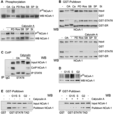
Regulation of gene expression represents a central issue in signal-regulated cellular responses. STAT6 is a critical mediator of IL-4 stimulated gene activation. To mediate this function, STAT6 recruits co-activator complexes. We have previously shown that STAT6 binds the PAS-B domain of the co-activator NCoA-1 via an LXXLL motif in its transactivation domain. Our recent finding that the PAS-B domain of NCoA-1 is also essential for co-activator complex formation points to an additional level of regulation of the co-activator assembly. In this study, we discovered that dephosphorylation of NCoA-1 is essential for the interaction with STAT6 and for IL-4-dependent transcriptional activation. PP2A dephosphorylates NCoA-1 and facilitates the activation of STAT6 target genes. Interestingly, simultaneous inhibition of phosphatase and cyclin-dependent kinases rescues the NCoA-1/STAT6 interaction. Moreover, arrest of cells at G1/S results in enhanced NCoA-1 phosphorylation. In summary, our results indicate that the interaction of NCoA-1 and STAT6 is dynamically regulated by the phosphatase PP2A and by cyclin-dependent kinases. This provides a mechanism for integrating transcriptional regulation by STAT6 with cell cycle progression.


Figures

Figure 1.
Figure 1.
Signaling pathways regulating STAT6/NCoA-1 interaction. 293T cells were transiently transfected with expression vectors encoding EGFP-STAT6 (100 ng) and hNCoA-1 (500 ng). Cells were treated with IL-4 (10 ng/ml) 24 h after transfection, kinase inhibitors PD98059 (PD, 50 µM), SB202190 (SB, 10 µM), SP600125 (SP, 200 nM), Staurosporin (St, 200 nM) or with the phosphatase inhibitor calyculin A (Ca, 10 nM) as indicated for 2 h. Cell extracts were prepared and co-immunoprecipitation (IP) was performed with an anti-GFP antibody (GFP), or an unspecific antibody (IgG). STAT6 bound NCoA-1 was analyzed by SDS–PAGE and western blotting with an anti-NCoA-1 antibody (left panel). Efficiency of the precipitation of GFP-STAT6 was proved by a GFP-specific antibody (right panel). As a control two percent of the cell lysates were analyzed in parallel (In).
Figure 2.
Figure 2.
Phosphatase inhibition induces STAT6 and NCoA-1 phosphorylation. (A) Ocadaic acid and calyculin A-dependent electromobility shift of STAT6 and NCoA-1. A549, Ramos and THP-1 cells were treated with 2 or 10 nM Ocadaic acid (OA) and calyculin A (Ca) for 2 h. Cells were lysed and electromobility of NCoA-1 and STAT6 were analyzed by SDS–PAGE and western blotting with NCoA-1 and STAT6 specific antibodies. (B) Phosphorylation of STAT6 and NCoA-1. 2 × 107 BJAB cells were metabolically labeled with 0.5 mCi P32. Cells were treated for 2 h with IL-4 (10 ng/ml); calyculin A (10 nM) alone or in combination; or with calyculin A after 30 min pretreatment with staurosporine (200 nM). STAT6 and NCoA-1 were immunoprecipitated and phosphorylation of the proteins was analyzed after SDS–PAGE by autoradiography. The immunoprecipitation was controlled in parallel by western blotting with specific antibodies (lower lane in both panels).
Figure 3.
Figure 3.
NCoA-1 dephosphorylation is essential for its interaction with STAT6. (A and B) Ramos B cells were stimulated with IL-4 (10 ng/ml) or treated with calyculin A (10 nM) or with calyculin A in combination with staurosporine (200 nM) for 2 h. Cells were lysed in NETN buffer and interaction of endogenous proteins were analyzed in GST pull-down experiments with coupled fusion proteins expressing the PAS domain of NCoA-1, GST-NCoA-1 Pas (A) or the transactivation domain of STAT6, GST-STAT6-TAD (B) or the activation domain 2 of the estrogen receptor, GST-ER E/F. Interaction of STAT6 (A) and NCoA-1 (B) was analyzed by western blotting with specific antibodies. (C) Treatment of the cells as in A. STAT6 was immunoprecipitated and co-percipitated NCoA-1 was detected after western blotting with specific antibodies. The efficiency of the STAT6 precipitation was examinated by reprobing with a STAT6 specific antibody. The amount of NCoA-1 in two percent of the cell lysates was analyzed in parallel (In). (D) Schematic model of the design and the results of the pull-down experiments performed in (A) (left panel) and in (B) (right panel).
Figure 4.
Figure 4.
NCoA-1 is dephosphorylated by PP2A. (A) Dephosphorylation of NCoA-1 by PP2A. 293T cells transfected with 3 µg of NCoA-1 expression vector were metabolically labeled 24 h after transfection with 0.5 mCi P32. Endogenous phosphatases were inactivated prior lysis by treatment with calyculin A (10 nM) for 10 min. For the labeling of BJAB cells, 6 × 107 cells were used. For the dephosphorylation assay PP2A immunoprecipitated out of unlabeled cells was used. As controls, a precipitation with an unspecific antibody (second lane) and a precipitation of PP2A under treatment with calyculin A (10 nM) (last lane) were used. The dephosphorylation reaction was carried out for 1 h at 37°C under shaking. NCoA-1 was immunoprecipitated out of the lysates and phosphorylation was visualized in autoradiography after SDS–PAGE (upper lanes). The amount of NCoA-1 was determined by western blotting (lower lanes). The phosphatase activity of the immunoprecipitated PP2A was determined in parallel (bar chart). (B) Interaction of PP2A with the NCoA family members. Endogenous NCoA-1, NCoA-2 and NCoA-3 were immunoprecipitated from BJAB cells by specific antibodies against the different NCoAs (NC). Coprecipitated PP2A was detected by western blotting with an antibody against the catalytic subunit of PP2A. As controls two percent of the cell lysates were analyzed in parallel (In) and unspecific binding was analyzed by precipitation with IgG. (C) Interaction of NCoA proteins with the A and C subunits of PP2A. Lysates of BJAB cells were incubated with coupeled GST-fusion proteins expressing the A subunit of PP2A (labeled A) or the C subunit of PP2A (labeled C) or with GST as control. Bound NCoA proteins were detected by western blotting with specific antibodies.
Figure 5.
Figure 5.
PP2A regulates the STAT6/ NCoA-1 interaction. (A) PP2A-dependent co-precipitation of NCoA-1 with STAT6. 293T cells transfected with 10 nM of two different siRNA against PP2A (siPP2A 1, siPP2A 2) or with an unspecific siRNA (scr) were transiently transfected 24 h later with plasmids encoding EGFP-STAT6 (1 µg) and NCoA-1 (2 µg) per 10-cm dish. One sample (scr + Ca) transfected with unspecific siRNA was treated with calyculin A (10 nM) for 2 h. Twenty-four hour later cells were lysed and STAT6 was precipitated by a GFP specific antibody. Bound NCoA-1 was detected after western blotting with specific antibody. Efficient precipitation of STAT6 was proven with a GFP specific antibody. As a control 2% of the cell lysates were analyzed in parallel (In). (B) PP2A-dependent interaction of NCoA-1 with STAT6 in pull-down experiments. Lysates of 293T cells transfected with specific or unspecific siRNAs as described in A were incubated with coupeled GST-STAT6-TAD fusion proteins or with GST as control. Bound NCoA-1 was detected after western blotting with specific antibody. As a control 10% of the cell lysates were analyzed in parallel (In).
Figure 6.
Figure 6.
PP2A regulates transcriptional activation by STAT6. (A) 293T cells were transiently transfected with a STAT6 reporter plasmid and expression plasmids for STAT6, β-galactosidase for normalization and GFP as transfection control. For the investigation of the estrogen receptor (ER) regulated transcription, cells were transfected with an ER reporter plasmid and expression plasmids for ER and the control plasmids for β-galactosidase and GFP. The cells were stimulated with IL-4 for 8 h or estrogen over night and treated with different concentrations of phosphatase inhibitors ocadaic acid (OA) and calyculin A (Ca). Cells were harvested and luciferase and β-galactosidase activities were determinated. The relatve luciferase activity was normalized to β-galactosidase. (B) 293T cells were transfected with unspecific or different siRNAs (10 nM) against PP2A or an expression vector encoding for HA-PP2A (50 ng) in addition to the STAT6 reporter and expression plasmids. One day later, the cells were stimulated with IL-4 and treated with 10 nM ocadaic acid (OA) or calyculin A (Ca) when indicated. Cells were harvested and luciferase and β-galactosidase activities were measured. (C) Ramos B cells were transfected with an unspecific siRNA or different siRNAs against PP2A (40 nM) by electroporation. Forty-eight hour post-transfection, the medium was changed to medium containing 1% FCS. After 8 h, the cells were stimulated with IL-4 for 24 h. One sample transfected with unspecific siRNA was treated in combination with calyculin A (2 nM). Total RNA was extracted and the expression level of Ig-epsilon and TARC were analyzed and normalized against 18S RNA. The results shown in (A), (B) and (C) are the average of three biological independent experiments.
Figure 7.
Figure 7.
NCoA-1 phosphorylation and its interaction potential for STAT6 is regulated by phosphatases and cell cycle-dependent kinases in an opposite direction. (A) Phosphorylation: 293T cells transiently transfected with NCoA-1 expression plasmids were metabolically labeled one day later in the presence of different inhibitors: Ocadaic acid (OA, 10 nM), calyculin A (Ca, 10 nM), PD98059 (PD, 50 µM), Roscovitin (Ros, 3 µM), SB202190 (SB, 10 µM), SP600125 (SP, 200 nM) and Staurosporin (St, 200 nM) for 2 h (upper panel). Cell labeling was also performed after 30 min pretreatment with calyculin A under combined treatment with calyculin A and different kinase inhibitors (lower panel). NCoA-1 was immunoprecipitated and the protein phosphorylation was determined by autoradiography. Immunoprecipitation was controlled by western blotting with an NCoA-1 specific antibody (lower lanes of both panels). (B) GST pull down: lysates of 293T cells treated with inhibitors as in (A) were incubated with GST-STAT6-TAD or with GST. Bound NCoA-1 was detected by western blotting. Ten percent of the cell lysates were analyzed in parallel as control (Input). (C) Co-immunoprecipitation: Ramos cells were treated with ocadaic acid (OA, 10 nM), calyculin A (Ca, 10 nM) or with calyculin A in combination with roscovitine (Ro, 3 µM) after 30 min pretreatment. STAT6 was precipitated out of the lysates and coprecipitated NCoA-1 was detected by western blotting. As a control 2% of the cell lysates were analyzed in parallel (Input). (D) 293T transiently transfected with NCoA-1 expression plasmids (1 µg) were arrested at different points of the cell cycle by overnight inhibitor treatment: late G1/S—double thymidine block (1 mM); early G1/S—hydroxyurea (0.5 mM) and G2—colcemide (100 nM). The cells were metabolically labeled with 0.1 mCi P32 and NCoA-1 was immunoprecipitated using an NCoA-1 specific antibody. Phosphorylation was detected by autoradiography (left panel, upper lane). Immunoprecipitation was controlled by western blotting (left panel, lower lanes). (E) For interaction assays cell lysates derived from cell cycle arrested 293T cells were incubated with GST or the GST-STAT6-TAD, bound NCoA-1 was detected by western blotting. As a control 10% of the cell lysates were analyzed in parallel (Input). One probe was derived from non arrested cells treated with calyculin A (10 nM) for 2 h for comparison (right lane). (F) To analyze if cell-cycle arrest can affect the inhibition of the NCoA-1/STAT6 interaction by calyculin A, cell-cycle arrested 293T cells were treated with calyculin A (10 nM) 2 h before lysate preparation and interaction assay.
Figure 8.
Figure 8.
Model for the regulated recruitment of NCoA-1 by STAT6. NCoA-1 can be phosphorylated by cdk2 and this prevents its interaction with STAT6. Dephosphorylation of NCoA-1 by PP2A is dominant and prerequisite for the recruitment of this co-activators to STAT6 responsive promoters where it facilitates the transcription by RNA polymerase II.

Similar articles

Cited by

References

    1. Nelms K, Keegan AD, Zamorano J, Ryan JJ, Paul WE. The IL-4 receptor: signaling mechanisms and biologic functions. Annu. Rev. Immunol. 1999;17:701–738. - PubMed
    1. Bruns HA, Kaplan MH. The role of constitutively active Stat6 in leukemia and lymphoma. Crit. Rev. Oncol. Hematol. 2006;57:245–253. - PubMed
    1. Hebenstreit D, Wirnsberger G, Horejs-Hoeck J, Duschl A. Signaling mechanisms, interaction partners, and target genes of STAT6. Cytokine Growth Factor Rev. 2006;17:173–188. - PubMed
    1. Torchia J, Glass C, Rosenfeld MG. Co-activators and co-repressors in the integration of transcriptional responses. Curr. Opin. Cell Biol. 1998;10:373–383. - PubMed
    1. Perissi V, Rosenfeld MG. Controlling nuclear receptors: the circular logic of cofactor cycles. Nat. Rev. Mol. Cell Biol. 2005;6:542–554. - PubMed

Publication types

MeSH terms