. 2011 Apr;92(Pt 4):733-43.
doi: 10.1099/vir.0.027052-0. Epub 2010 Dec 9.

Syndecan-1 and syndecan-2 play key roles in herpes simplex virus type-1 infection

Affiliations

Syndecan-1 and syndecan-2 play key roles in herpes simplex virus type-1 infection

Sarolta Bacsa et al. J Gen Virol. 2011 Apr.

Abstract

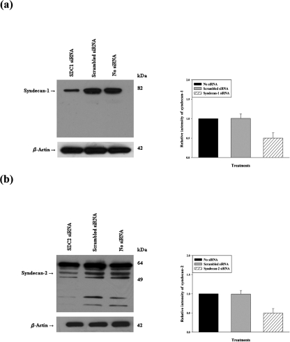
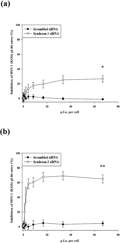
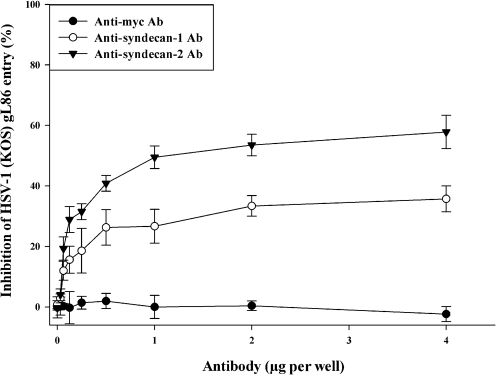
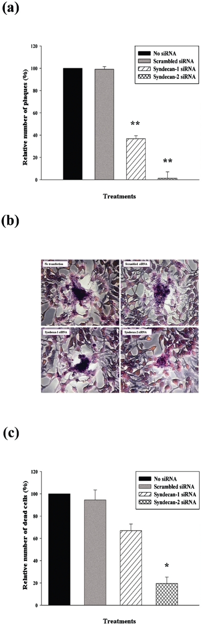
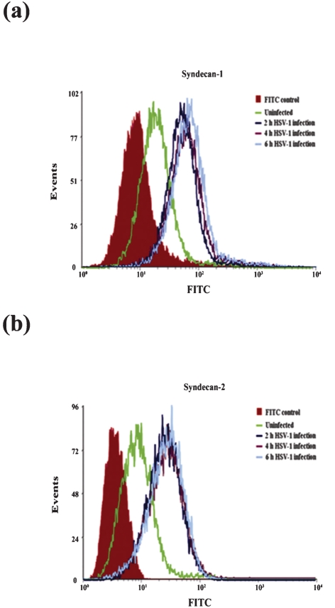
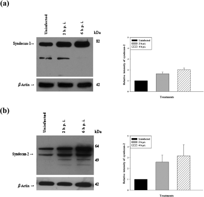
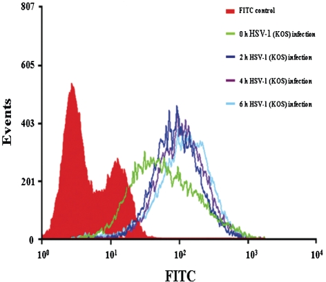
Herpes simplex virus type 1 (HSV-1) is an important human pathogen and a leading cause of infectious blindness in the developed world. HSV-1 exploits heparan sulfate proteoglycans (HSPG) for attachment to cells. While the significance of heparan sulphate (HS) moieties in HSV-1 infection is well established, the role of specific proteoglycan core proteins in the infection process remains poorly understood. The objective of this study was to assess the roles of syndecan-1 and syndecan-2 core proteins in HSV-1 infection, both of which are expressed by many HSV-1 target cell types. Our results demonstrate that syndecan-1 and syndecan-2 gene silencing by RNA interference reduces HSV-1 entry, plaque formation and facilitates cell survival. Furthermore, HSV-1 infection increases syndecan-1 and syndecan-2 protein synthesis and a resultant increase in cell surface expression of HS. Our observations suggest that changes in syndecan-1 and syndecan-2 expression levels may be related to active viral infection. Taken together, our findings provide new insights into HSPG functions during HSV-1 entry and spread.


Figures

Fig. 1.
Fig. 1.
Western blot analysis of syndecan-1 and syndecan-2 protein expression after siRNA downregulation. Protein expression of syndecan-1 and syndecan-2 measured in a sample of HeLa cells mock treated (no transfection) or treated with scrambled siRNA, syndecan-1 siRNA or syndecan-2 siRNA for 48 h. Representative Western blots showed knockdown of syndecan-1 (a) or syndecan-2 (b) after siRNA downregulation. Densitometric analysis revealed 50 % reduction in the signal intensity of both syndecan-1 and syndecan-2. β-Actin protein expression was measured as the loading control. Representative blots from three independent experiments are shown. The results are expressed as means±sd values from three independent experiments.
Fig. 2.
Fig. 2.
Downregulation of syndecan-1 and syndecan-2 inhibits HSV-1 entry into HeLa cells. HSV-1 entry was analysed in HeLa cells mock treated (no transfection) or transfected with scrambled siRNA, syndecan-1 siRNA or syndecan-2 siRNA. After siRNA transfection (48 h), cells were inoculated with a serial dilution of β-galactosidase-expressing recombinant HSV-1 (KOS) gL86 virus for 6 h. The soluble substrate ONPG was added and enzymic activity was measured. Downregulation of both syndecan-1 (Fig. 2a) and syndecan-2 (Fig. 2b) inhibits HSV-1 (KOS) gL86 entry into HeLa cells although syndecan-2 siRNA has more significant effect. Per cent inhibition of HSV-1(KOS) gL86 entry by syndecan-1 siRNA or syndecan-2 siRNA treatment was calculated relative to mock-treated (no transfection) cells. Scrambled siRNA-transfected cells were used as negative control. Each value shown is the mean of six determinations (±sd) (*P<0.05, **P<0.0001).
Fig. 3.
Fig. 3.
Syndecan-1- or syndecan-2-specific antibody treatment blocked HSV-1(KOS) gL86 entry into HeLa cells. Cells were incubated with a serial dilution of syndecan-1-specific pAbs, syndecan-2-specific pAbs, or control anti-myc mAb for 30 min. Cells were then inoculated with a constant dose of β-galactosidase-expressing recombinant HSV-1(KOS) gL86 (m.o.i. of 10) for 2 h. The soluble substrate ONPG was added and enzymic activity was measured. Per cent inhibition of HSV-1(KOS) gL86 entry by syndecan-1 pAbs or syndecan-2 pAbs treatment was calculated relative to anti-myc mAb-treated cells. Each value shown is the mean of four determinations (±sd) (P<0.05).
Fig. 4.
Fig. 4.
Downregulation of syndecan-1 and syndecan-2 affects HSV-1 plaque formation, size and enhances cell survival in HeLa cells. Cells were either mock treated (no transfection), or treated with scrambled siRNA, syndecan-1 siRNA or syndecan-2 siRNA. Post-transfection cells (48 h) were infected with HSV-1(KOS) (m.o.i. of 0.01). (a) Post-infection infectivity at 72 h was measured by the number of p.f.u. Relative number of plaques was computed relative to mock-treated (no transfection) samples. Significant decreases of number of plaques were seen in both syndecan-1 siRNA- and syndecan-2 siRNA-transfected HeLa cells. Plaques that consisted of 15 or more nuclei were counted. Results are means±sd of four independent experiments conducted in triplicate (**P<0.0001). (b) Morphological appearance of Giemsa-stained HSV-1 (KOS) plaques 72 h post-infection. In syndecan-1 siRNA- and syndecan-2 siRNA-treated HeLa cells smaller plaques were observed compared with the plaques in mock-treated or scrambled siRNA-treated cells. Magnification, ×40. (c) Cytotoxicity was measured after 120 h HSV-1 (KOS) infection. Relative number of dead cells was calculated relative to mock-treated samples. Significant decreases of cytotoxicity were observed in both syndecan-1 siRNA- and syndecan-2 siRNA-treated cells; however, only the effect of syndecan-2 siRNA treatment was statistically significant. Results are means±sd of four independent experiments conducted in triplicate (*P<0.05).
Fig. 5.
Fig. 5.
HSV-1 infection of host cell enhances syndecan-1 and syndecan-2 cell surface expression in HeLa cells. Cells were infected with a constant dose of HSV-1 (KOS) (m.o.i. of 10) for 2, 4 or 6 h. Syndecan-1 and syndecan-2 cell surface expression was then detected by FACS analysis. Enhanced syndecan-1 (a) and syndecan-2 (b) cell surface expression was detected when HeLa cells were infected with HSV-1. Mock-infected FITC stained cells were used as background control. Results are representative of three independent experiments.
Fig. 6.
Fig. 6.
Western blot analysis of syndecan-1 and syndecan-2 protein expression after HSV-1 (KOS) infection. HeLa cells were infected with a constant dose of HSV-1 (KOS) (m.o.i. of 10) for 2 and 6 h. Cell lysates were then blotted against syndecan-1 and syndecan-2. According to the densitometric analysis syndecan-1 (a) and syndecan-2 (b) were more strongly expressed at 2 and 6 h post-infection compared with the uninfected cells in HeLa cells. Relative intensity of HSV-1(KOS) virus-infected bands expressed as a ratio relative to the mock-infected sample is shown. β-Actin protein expression was measured as loading control. The results are expressed as means±sd values from three independent experiments.
Fig. 7.
Fig. 7.
Flow cytometry analysis of HS expression on HeLa cells infected with HSV-1. HeLa cells were infected with HSV-1(KOS) (m.o.i. of 10) at 37 °C for 2, 4 or 6 h. HS cell surface expression was then detected by FACS analysis. In HSV-1-infected cells, HS cell surface expression was enhanced compared with uninfected cells. Untreated FITC stained HeLa cells were used as background control.

References

    1. Akhtar, J., Tiwari, V., Oh, M. J., Kovacs, M., Jani, A., Kovacs, S. K., Valyi-Nagy, T. & Shukla, D. (2008). HVEM and nectin-1 are the major mediators of herpes simplex virus 1 (HSV-1) entry into human conjunctival epithelium. Invest Ophthalmol Vis Sci 49, 4026–4035. - PMC - PubMed
    1. Anastasiadou, E., Vaeth, S., Cuomo, L., Boccellato, F., Vincenti, S., Cirone, M., Presutti, C., Junker, S., Winberg, G. & Frati, L. (2009). Epstein–Barr virus infection leads to partial phenotypic reversion of terminally differentiated malignant B cells. Cancer Lett 284, 165–174. - PubMed
    1. Atanasiu, D., Whitbeck, J. C., Cairns, T. M., Reilly, B., Cohen, G. H. & Eisenberg, R. J. (2007). Bimolecular complementation reveals that glycoproteins gB and gH/gL of herpes simplex virus interact with each other during cell fusion. Proc Natl Acad Sci U S A 104, 18718–18723. - PMC - PubMed
    1. Barth, H., Schafer, C., Adah, M. I., Zhang, F., Linhardt, R. J., Toyoda, H., Kinoshita-Toyoda, A., Toida, T., Van Kuppevelt, T. H. & other authors (2003). Cellular binding of hepatitis C virus envelope glycoprotein E2 requires cell surface heparan sulfate. J Biol Chem 278, 41003–41012. - PubMed
    1. Beauvais, D. M., Burbach, B. J. & Rapraeger, A. C. (2004). The syndecan-1 ectodomain regulates αvβ3 integrin activity in human mammary carcinoma cells. J Cell Biol 167, 171–181. - PMC - PubMed

Publication types

LinkOut - more resources