. 2011 Jan 1;22(1):44-53.
doi: 10.1091/mbc.E10-08-0720. Epub 2010 Dec 9.

Actin cables and the exocyst form two independent morphogenesis pathways in the fission yeast

Affiliations

Actin cables and the exocyst form two independent morphogenesis pathways in the fission yeast

Felipe O Bendezú et al. Mol Biol Cell. .

Abstract

Cell morphogenesis depends on polarized exocytosis. One widely held model posits that long-range transport and exocyst-dependent tethering of exocytic vesicles at the plasma membrane sequentially drive this process. Here, we describe that disruption of either actin-based long-range transport and microtubules or the exocyst did not abolish polarized growth in rod-shaped fission yeast cells. However, disruption of both actin cables and exocyst led to isotropic growth. Exocytic vesicles localized to cell tips in single mutants but were dispersed in double mutants. In contrast, a marker for active Cdc42, a major polarity landmark, localized to discreet cortical sites even in double mutants. Localization and photobleaching studies show that the exocyst subunits Sec6 and Sec8 localize to cell tips largely independently of the actin cytoskeleton, but in a cdc42 and phospholipid phosphatidylinositol 4,5-bisphosphate (PIP₂)-dependent manner. Thus in fission yeast long-range cytoskeletal transport and PIP₂-dependent exocyst represent parallel morphogenetic modules downstream of Cdc42, raising the possibility of similar mechanisms in other cell types.


Figures

FIGURE 1:
FIGURE 1:
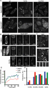
Polarized actin cables and microtubules are not required for polarized cell growth. (A–E) Wild-type, for3Δ fus1Δ cdc12112 and myo51Δ myo52Δ mutant cells stained with TRITC-lectin (green; all panels) and calcofluor (red; right panels). Cells were grown at 25°C (A, D, E) or 36°C (B, C) or were treated with MBC for 5 h as indicated (C and E). Clearing of fluorescent lectin at cell ends shows regions of growth (arrowheads). (F) GFP-Bgs1 and GFP-Syb1 signals in wild-type, for3Δ, and myo51Δ myo52Δ grown with or without MBC for 30 min. Arrowheads indicate enrichment of fusion proteins. Boxed regions are shown enlarged in the bottom row. (G) Recovery of GFP-Bgs1 to bleached tips in wild-type and myo51Δ myo52Δ in the presence of MBC. (H) Average recovery of GFP-Bgs1 to bleached tips (n = 15–20). GFP-Bgs1 shows a similar half-time of recovery of <5 s and immobile fraction of 60–66% in all strains. (I) Secretion of acid phosphatase in wild-type, myo51Δ myo52Δ, and for3Δ cells. Bars, 5 μm.
FIGURE 2:
FIGURE 2:
The exocyst localizes to cell tips in absence of actin cables. (A) Sec6-GFP and Sec8-GFP signals in wild-type, for3Δ, and myo52Δ cells. (B) Recovery of Sec6-GFP to bleached tips in the presence of LatA or DMSO. Actin depolymerization was confirmed by coimaging of cells expressing the F-actin binding protein GFP-CHD (left panels). (C) Sec6-GFP in wild-type treated with LatA for 5 and 30 min. Bottom panels show recovery of side wall staining bleached in inset. Arrowheads, arrows, and open triangles indicate tip, cell side, and division site localization, respectively. Bars, 5 μm. (D) Average recovery of Sec6-GFP to bleached tips (n = 15). Control, for3Δ, and LatA-treated cells show 5-, 5-, and 10-s half-time of recovery and 35%, 35%, and 60% immobile fractions, respectively. (E) Frequency of excellent (>60%), good (40–60%), medium (20–40%), or poor (0–20%) recovery to bleached tips by averaging recovery from 40 to 80 s.
FIGURE 3:
FIGURE 3:
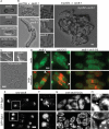
Loss of both actin cable machinery and the exocyst results in failure to polarize growth. (A) Germinated sec6Δ, sec8Δ, sec6Δ sec8Δ, and sec8Δ exo70Δ spores on YE5S at 25°C. (B) Cells with indicated genotype from cross of exo70Δ × sec81 or myo52Δ × sec81. (C) exo70Δ, for3Δ, and for3Δ exo70Δ cells. (D) sec81, cdc3313, and sec81 cdc3313 cells stained with TRITC-lectin (green; all panels) and calcofluor (red; bottom panels). Note clearing of lectin staining at cell ends of single mutants (arrowheads) but absence of clearing and patchy appearance in double mutant cells. (E–H) GFP-Bgs1 and GFP-Syb1 signals in nmt-sec8 and for3Δ nmt-sec8 cells depleted for Sec8. (F, H) Enlargements of insets marked in (E, G). Arrowheads indicate cortical and arrows indicate subcortical polar accumulation of fusion proteins. Bars, 5 μm.
FIGURE 4:
FIGURE 4:
Cdc42 activity is independent of actin cable and exocyst and is necessary for exocyst localization. (A) CRIB-GFP in wild-type, for3Δ, nmt-sec8, and for3Δ nmt-sec8 cells depleted for Sec8. (B) RFP-Bgs4 (red) and CRIB-GFP (green) in wild-type, cdc3313, sec81, and cdc3313 sec81 cells grown for 90 min at 36°C. (C) Sec6-GFP and Sec8-GFP in wild-type (left panels), cdc421625 (middle panels), and cdc42879 mutant cells. CRIB-GFP in cdc421625 mutant is also shown as labeled. (D) Localization of CRIB-GFP and For3–3GFP in wild-type cells treated with LatA for 5 and 30 min. Arrows indicate polar and arrowheads lateral localization of fusion proteins. Bars, 5 μm.
FIGURE 5:
FIGURE 5:
The localization of the exocyst, but not of active Cdc42 or Myo52, is dependent on PIP2. (A) Sec6-GFP, Sec8-GFP, CRIB-GFP, and Myo52–3GFP signals in wild-type and its31 mutant cells grown at 36°C in EMM for 90 min. (B) its31 sec81 and its31 for3Δ double-mutant and control cells. Bar, 5 μm.
FIGURE 6:
FIGURE 6:
Model for parallel morphogenesis pathways under the control of Cdc42. Cdc42 sets up cell polarity and regulates two parallel morphogenetic modules for polarized cell growth, the formin-dependent actin cable module and the exocyst module, contributing to transport and tethering of exocytic vesicles, respectively. The exocyst is also controlled by PIP2 levels at the plasma membrane. Direct links between Cdc42, PIP2, and the exocyst are as yet unknown.

References

    1. Ayscough KR, Stryker J, Pokala N, Sanders M, Crews P, Drubin DG. High rates of actin filament turnover in budding yeast and roles for actin in establishment and maintenance of cell polarity revealed using the actin inhibitor latrunculin-A. J Cell Biol. 1997;137:399–416. - PMC - PubMed
    1. Balasubramanian MK, Helfman DM, Hemmingsen SM. A new tropomyosin essential for cytokinesis in the fission yeast S. pombe. Nature. 1992;360:84–87. - PubMed
    1. Balasubramanian MK, Hirani BR, Burke JD, Gould KL. The Schizosaccharomyces pombe cdc3+ gene encodes a profilin essential for cytokinesis. J Cell Biol. 1994;125:1289–1301. - PMC - PubMed
    1. Boyd C, Hughes T, Pypaert M, Novick P. Vesicles carry most exocyst subunits to exocytic sites marked by the remaining two subunits, Sec3p and Exo70p. J Cell Biol. 2004;167:889–901. - PMC - PubMed
    1. Chalkia D, Nikolaidis N, Makalowski W, Klein J, Nei M. Origins and evolution of the formin multigene family that is involved in the formation of actin filaments. Mol Biol Evol. 2008;25:2717–2733. - PMC - PubMed

Publication types

MeSH terms