Review
. 2010 Dec 10;107(12):1428-44.
doi: 10.1161/CIRCRESAHA.110.227405.

Myocardial lineage development

Affiliations
Review

Myocardial lineage development

Sylvia M Evans et al. Circ Res. .

Abstract

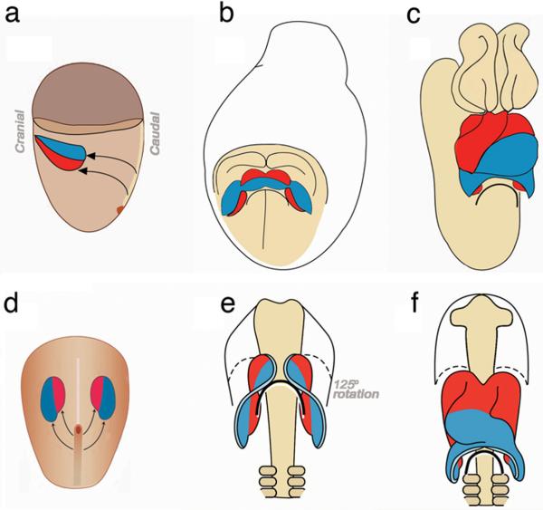
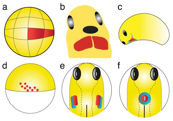
The myocardium of the heart is composed of multiple highly specialized myocardial lineages, including those of the ventricular and atrial myocardium, and the specialized conduction system. Specification and maturation of each of these lineages during heart development is a highly ordered, ongoing process involving multiple signaling pathways and their intersection with transcriptional regulatory networks. Here, we attempt to summarize and compare much of what we know about specification and maturation of myocardial lineages from studies in several different vertebrate model systems. To date, most research has focused on early specification, and although there is still more to learn about early specification, less is known about factors that promote subsequent maturation of myocardial lineages required to build the functioning adult heart.


Figures

Figure 1
Figure 1
Schematic representation of heart tube formation in mouse (a, b, c) and chick (d, e, f). First heart progenitors are shown in blue and second heart progenitors in red. (a,d) Fate mapping studies have defined the locations of chick and mouse cardiac progenitors in the heart fields. (a) Lateral view of an E6.5 mouse embryo. (d) Dorsal view of an HH5 chick embryo. (b,e) Fusion and differentiation of the heart fields is more rapid in the mouse leading to a cardiac crescent that is not seen in the chick. Second heart progenitors are located medially in both chick and mouse heart fields but quickly change positions to cranial as the heart fields converge on the midline. (b) Ventral view of an E8.5 mouse embryo showing the cardiac crescent and its relationship with the anterior intestinal portal (curved black line). (e) Ventral view of an HH8 chick embryo showing convergence of the heart fields in the ventral midline and how the first and second heart field progenitors have rotated their position from medial-lateral to craniocaudal. (c,f) Second heart progenitors are gradually added to the elongating cardiac tube. (c) Ventral view of an E9.5 mouse embryo. (f) Ventral view of an HH12 chick embryo. The caudal second heart progenitors are shifted by formation of the foregut pocket and anterior intestinal portal (curved black line) to cranial, thus putting them in place to contribute to the outflow pole. Some of the second heart field progenitors are also added to the venous pole: parts of the atrium and atrial septum but these are incorporated later than the stages shown, hence no red cells are seen at these stages in the venous pole. The proximal and distal outflow myocardium is added over an extended period of time (reprinted from with permission).
Figure 2
Figure 2
Schematic representation of key stages in heart tube formation in frog (a, b, c) and fish (d, e, f). (a, d) Fate mapping studies have defined the locations of frog and fish cardiac progenitors prior to gastrulation. (a) Lateral view of a 32-cell stage Xenopus embryo, dorsal to the right, indicating the blastomeres (red) that will give rise to descendants in the adult heart. (d) Lateral view of a zebrafish embryo at the 40% epiboly stage, dorsal to the right, indicating the approximate locations of the blastomeres (red) that will give rise to the myocardium. (b, e) Fate maps and the expression patterns of molecular markers have demonstrated the locations of the bilateral heart fields in both frog and fish. (b) Anterior-ventral view of a Xenopus mid-tadpole, anterior to the top, just prior to the fusion of the heart fields (red) at the ventral midline. Cement gland is labeled in black and eyes are labeled in grey. Note: In Xenopus, unlike zebrafish, the atrial and ventricular precursor populations are not distinct populations but rather the precursors are intermingled until very late in heart development. (e) Dorsal view of a zebrafish embryo at the 7-somite stage, anterior to the top, indicating the locations of ventricular myocardial progenitors (red) and atrial myocardial progenitors (blue) prior to cardiac fusion. Notochord is labeled in black and eyes are labeled in grey. (c. f) Expression patterns of molecular markers have indicated the existence of discrete myocardial subpopulations as the heart tube forms. (c) Lateral view of a Xenopus tadpole at stage 25, anterior to the left, after fusion of the cardiac progenitors at the midline. Molecular markers distinguish cardiac populations expressing Tbx1 and Isl-1 (lavender) from those that do not (green). (f) Dorsal view of a zebrafish embryo at the 21-somite stage, anterior to the top, indicating the locations of ventricular cardiomyocytes (red) and atrial cardiomyocytes (blue) within the cardiac cone.

Similar articles

Cited by

References

    1. Vincent SD, Buckingham ME. How to make a heart: the origin and regulation of cardiac progenitor cells. Curr Top Dev Biol. 2010;90:1–41. - PubMed
    1. Thomas PQ, Brown A, Beddington RS. Hex: a homeobox gene revealing peri-implantation asymmetry in the mouse embryo and an early transient marker of endothelial cell precursors. Development. 1998;125:85–94. - PubMed
    1. Tam PPL, Parameswaran M, Kinder SJ, Weinberger RP. The allocation of epiblast cells to the embryonic heart and other mesodermal lineages: The role of ingression and tissue movement during gastrulation. Development. 1997;124:1631–1642. - PubMed
    1. Garcia-Martinez V, Schoenwolf GC. Primitive-streak origin of the cardiovascular system in avian embryos. Dev. Biol. 1993;159:706–719. - PubMed
    1. Bellairs R. The primitive streak. Anat Embryol (Berl) 1986;174:1–14. - PubMed

Publication types