Clinical Trial
. 2011 Jan 1;52(1):1-7.
doi: 10.1093/cid/ciq015.

Potent CD8+ T-cell immunogenicity in humans of a novel heterosubtypic influenza A vaccine, MVA-NP+M1

Affiliations
Clinical Trial

Potent CD8+ T-cell immunogenicity in humans of a novel heterosubtypic influenza A vaccine, MVA-NP+M1

Tamara K Berthoud et al. Clin Infect Dis. .

Abstract

Background: Influenza A viruses cause occasional pandemics and frequent epidemics. Licensed influenza vaccines that induce high antibody titers to the highly polymorphic viral surface antigen hemagglutinin must be re-formulated and readministered annually. A vaccine providing protective immunity to the highly conserved internal antigens could provide longer-lasting protection against multiple influenza subtypes.

Methods: We prepared a Modified Vaccinia virus Ankara (MVA) vector encoding nucleoprotein and matrix protein 1 (MVA-NP+M1) and conducted a phase I clinical trial in healthy adults.

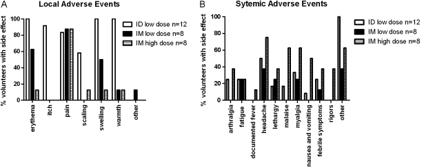
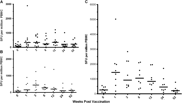
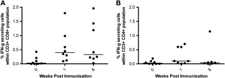
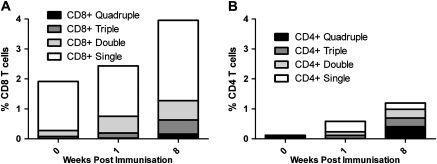
Results: The vaccine was generally safe and well tolerated, with significantly fewer local side effects after intramuscular rather than intradermal administration. Systemic side effects increased at the higher dose in both frequency and severity, with 5 out of 8 volunteers experiencing severe nausea/vomiting, malaise, or rigors. Ex vivo T-cell responses to NP and M1 measured by IFN-γ ELISPOT assay were significantly increased after vaccination (prevaccination median of 123 spot-forming units/million peripheral blood mononuclear cells, postvaccination peak response median 339, 443, and 1443 in low-dose intradermal, low-dose intramuscular, and high-dose intramuscular groups, respectively), and the majority of the antigen-specific T cells were CD8(+).

Conclusions: We conclude that the vaccine was both safe and remarkably immunogenic, leading to frequencies of responding T cells that appear to be much higher than those induced by any other influenza vaccination approach. Further studies will be required to find the optimum dose and to assess whether the increased T-cell response to conserved influenza proteins results in protection from influenza disease.


Figures

Figure 1.
Figure 1.
Local and systemic adverse events recorded after vaccination. Black: group 1 (n = 12). White: group 2 (n = 8). Striped: group 3 (n = 8). (A) Local adverse events. Significantly less (P < .05, Fisher's exact test) erythema, itch, swelling, and warmth at the injection site were detected in those receiving intramuscular vaccine than those vaccinated intradermally, regardless of the vaccine dose. Significantly less scaling was recorded in the low-dose compared with the high-dose intramuscular group. (B) Systemic adverse events. No significant differences in systemic adverse events were reported by the volunteers receiving the low-dose vaccine by either route, but there was a significant increase in malaise, nausea/vomiting, and rigors in the group receiving the high dose (P < .05, Fisher's exact test). Severe adverse events only occurred in the high-dose group, with 2 volunteers reporting severe pain at the injection site, 1 reporting malaise, 1 vomiting, 2 rigors, and 1 sweating. All severe adverse events resolved within 48 hours of vaccination, apart from 1 volunteer reporting severe pain at the injection site on the 3 days following vaccination. The majority of mild and moderate adverse events also took place within 48 hours of vaccination, although mild erythema at the injection site lasted for up to 49 days for those receiving intradermal vaccination.
Figure 2.
Figure 2.
Ex vivo IFN-γ ELISPOT responses to the vaccine insert. Median with individual ex vivo IFN-γ ELISPOT responses from vaccinated volunteers at baseline (week 0), and weeks 1, 3, 8, 12, 24, and 52 weeks after immunization. (A) group 1; (B) group 2; (C) group 3. Wilcoxon signed rank test was used to determine significant differences in the post- and prevaccination time points. (A) week 1, P = .0059; week 3, P = .0098; week 8, P = .0078; week 12, P = .0049. (B) week 1, P = .0313; week 3, P = .0313. (C) week 1, P = .0078; week 3, P = .0078; week 8, P = .0078; week 12, P = .0078; week 24, P = .023. Significant differences were detected between groups 2 (B) and 3 (C) at all postvaccination time points apart from week 52 (Mann-Whitney U test: week 1, P = .006; week 3, P = .04; week 8, P = .01; week 12, P = .02; week 24, P = .012).
Figure 3.
Figure 3.
CD3+CD4+ and CD3+CD8+ IFN-γ responses to vaccine insert as measured by intracellular cytokine staining. Intracellular IFN-γ responses after background subtraction in (A) CD3+CD8+ and (B) CD3+CD4+ cell populations stimulated with 1 pool of peptides spanning the complete NP+M1 vaccine insert. Volunteers in group 3 were tested at weeks 0, 1, and 8. Median % IFN-γ+ within CD3+CD8+ cells at week 1 = .4% and week 8 = .33%; median % IFN-γ+ cells within CD3+CD4+ population at week 1 = .098% and week 8 = .039%.
Figure 4.
Figure 4.
IFN-γ, IL-2, TNF-α, and CD107a multifunctional cells detected by ICS in CD3+CD8+ and CD3+CD4+ populations. Mean percentage of quadruple (black), triple (dark gray), double (light gray), and single (white) functional cells detected within the CD8+ (A) and CD4+ (B) populations. Within the CD8+ population, the most frequently detected triple positive cells were CD107a+IFN-γ+TNF-α+; the most frequently detected double positive cells were CD107a+TNF-α+; and CD107a+ cells were the most frequently detected single positive cells. Within the CD4+ cells, the most frequently detected triple positive cells were CD107a+IFN-γ+IL-2+; the most frequently detected double positive cells were IFN-γ+IL-2+; and TNF-α+ cells were the most frequently detected single positive cells. At all time points the frequency of antigen-specific cytokine positive cells was greater in the CD8+ population (week 0, CD8+ = 1.92% and CD4+ = .12%; week 1, CD8+ = 2.43% and CD4+ = .58%; week 8, CD8+ = 3.96% and CD4+ = 1.19%).

Comment in

Similar articles

Cited by

References

    1. Centers for Disease Control and Prevention (CDC) Interim within-season estimate of the effectiveness of trivalent inactivated influenza vaccine–Marshfield, Wisconsin, 2007-08 influenza season. MMWR Morb Mortal Wkly Rep. 2008;57:393–398. - PubMed
    1. de Jong JC, Beyer WE, Palache AM, Rimmelzwaan GF, Osterhaus AD. Mismatch between the 1997/1998 influenza vaccine and the major epidemic A(H3N2) virus strain as the cause of an inadequate vaccine-induced antibody response to this strain in the elderly. J Med Virol. 2000;61:94–99. - PubMed
    1. Galli G, Medini D, Borgogni E, et al. Adjuvanted H5N1 vaccine induces early CD4+ T cell response that predicts long-term persistence of protective antibody levels. Proc Natl Acad Sci U S A. 2009;106:3877–3882. - PMC - PubMed
    1. Nolan TM, Richmond PC, Skeljo MV, et al. Phase I and II randomised trials of the safety and immunogenicity of a prototype adjuvanted inactivated split-virus influenza A (H5N1) vaccine in healthy adults. Vaccine. 2008;26:4160–4167. - PubMed
    1. Breathnach CC, Clark HJ, Clark RC, Olsen CW, Townsend HG, Lunn DP. Immunization with recombinant modified vaccinia Ankara (rMVA) constructs encoding the HA or NP gene protects ponies from equine influenza virus challenge. Vaccine. 2006;24:1180–1190. - PubMed

Publication types

MeSH terms