. 2011 Feb;193(4):887-95.
doi: 10.1128/JB.00730-10. Epub 2010 Dec 10.

FAD binding by ApbE protein from Salmonella enterica: a new class of FAD-binding proteins

Affiliations

FAD binding by ApbE protein from Salmonella enterica: a new class of FAD-binding proteins

Jeffery M Boyd et al. J Bacteriol. 2011 Feb.

Abstract

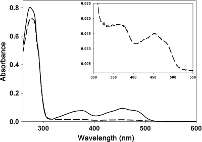
The periplasmic protein ApbE was identified through the analysis of several mutants defective in thiamine biosynthesis and was implicated as having a role in iron-sulfur cluster biosynthesis or repair. While mutations in apbE cause decreased activity of several iron-sulfur enzymes in vivo, the specific role of ApbE remains unknown. Members of the AbpE family include NosX and RnfF, which have been implicated in oxidation-reduction associated with nitrous oxide and nitrogen metabolism, respectively. In this work, we show that ApbE binds one FAD molecule per monomeric unit. The structure of ApbE in the presence of bound FAD reveals a new FAD-binding motif. Protein variants that are nonfunctional in vivo were generated by random and targeted mutagenesis. Each variant was substituted in the environment of the FAD and analyzed for FAD binding after reconstitution. The variant that altered a key tyrosine residue involved in FAD binding prevented reconstitution of the protein.


Figures

FIG. 1.
FIG. 1.
rApbE is a flavoprotein. The UV-visible absorption spectra of 15 μM rApbE before (dashed curve) and after (solid curve) reconstitution with FAD indicate that rApbE is a flavoprotein. (Inset) Magnification showing the relevant region of the UV-visible spectrum of the as-isolated rApbE.
FIG. 2.
FIG. 2.
FAD is released from purified rApbE. Authentic FAD (A) or flavin isolated from heat-denatured rApbE (B) was separated by LC. The peak eluting 6.94 min after injection was analyzed by electrospray ionization using a tandem in-line TOF mass spectrometer, and the mass/charge ratios are expressed as atomic mass units (amu) (insets).
FIG. 3.
FIG. 3.
rApbE protein is a dimer in solution. A plot of the log of known molecular mass markers (filled circles) versus the time taken for samples to elute from the column (Te/To) was used to determine the approximate size of the rApbE protein. The line shown is the fit to equation 1, which was used to conclude that FAD-reconstituted rApbE protein (empty square) has a Stokes radius constant with a molecular mass of approximately 52 kDa. The standards used were thyroglobulin (bovine, 670,000 Da), gamma globulin (bovine, 158,000 Da), ovalbumin (chicken, 44,000 Da), myoglobin (horse, 17,000 Da), and vitamin B12 (1,350 Da). (Inset) ApbE (0.5 mg) protein as isolated or after reconstitution with FAD (as shown) migrated as a single predominant species. The gel filtration conditions are as follows: solid phase, Superose 6; mobile phase, 50 mM Tris-HCl, pH 7.6, 200 mM NaCl; flow rate, 0.1 ml/min.
FIG. 4.
FIG. 4.
(A) Ribbon diagram of the rApbE dimer showing one FAD molecule bound per rApbE monomer related to the primary sequence. (B) Ribbon diagram of the rApbE monomer with FAD bound. The FAD is shown as a sphere representation. The phosphates are shown in magenta. (C) Ribbon diagrams of the rApbE monomer. The α helices are shown in purple, and the β sheets are shown in green. The residues of the FAD-binding site are shown in blue stick representation and are indicated on the sequence. The residues comprising the dimer interface are shown in salmon as a stick representation and are indicated on the sequence. Residues Ser76, Thr184, and Gly255 are indicated by black dots; and Tyr78, which was targeted for mutagenesis, is indicated by a black star.
FIG. 5.
FIG. 5.
(A) Stereo view of the FAD-binding site in the rApbE monomer. Residues interacting with the FAD isoalloxazine ring (B), phosphates (C), and the adenine (D) are shown in line angle representations.
FIG. 6.
FIG. 6.
The ApbEY78A variant is not functional in vivo. Growth of S. enterica strain DM9722 with pJMB129 (ApbEY78A; squares), pApbE1 (ApbE; circles), or pSU19 (empty vector; triangles) in minimal glucose medium supplemented with adenine is shown. Growth of the strains is shown as the absorbance at 650 nm over time in the presence of 100 nM thiamine (filled symbols) and with no thiamine added (open symbols). Variant proteins accumulated to levels that were indistinguishable from those of the wild-type protein by Western blot analysis (see text).
FIG. 7.
FIG. 7.
The rApbEY78A variant is defective in FAD binding. The UV-visible absorption spectra of wild-type rApbE (12 μM; solid line) and the Y78A rApbE variant (12 μM; dashed line) are shown. The proteins were reconstituted as described in the text. Without reconstitution, no absorbance in the 300- to 500-nm range was seen with the rApbEY78A variant.

References

    1. Agar, J. N., et al. 2000. IscU as a scaffold for iron-sulfur cluster biosynthesis: sequential assembly of [2Fe-2S] and [4Fe-4S] clusters in IscU. Biochemistry 39:7856-7862. - PubMed
    1. Backiel, J., et al. 2008. Covalent binding of flavins to RnfG and RnfD in the Rnf complex from Vibrio cholerae. Biochemistry 47:11273-11284. - PMC - PubMed
    1. Balch, W. E., and R. S. Wolfe. 1976. New approach to the cultivation of methanogenic bacteria: 2-mercaptoethanesulfonic acid-dependent growth of Methanobacterium ruminantium in a pressurized atmosphere. Appl. Environ. Microbiol. 32:781-791. - PMC - PubMed
    1. Bartolomé, B., Y. Jubete, E. Martinez, and F. de la Cruz. 1991. Construction and properties of a family of pACYC184-derived cloning vectors compatible with pBR322 and its derivatives. Gene 102:75-78. - PubMed
    1. Beck, B. J., and D. M. Downs. 1998. The apbE gene encodes a lipoprotein involved in thiamine synthesis in Salmonella typhimurium. J. Bacteriol. 180:885-891. - PMC - PubMed

Publication types

MeSH terms

Associated data

LinkOut - more resources