. 2010 Dec 28;107(52):22505-10.
doi: 10.1073/pnas.1007626107. Epub 2010 Dec 10.

Structural basis for active site closure by the poliovirus RNA-dependent RNA polymerase

Affiliations

Structural basis for active site closure by the poliovirus RNA-dependent RNA polymerase

Peng Gong et al. Proc Natl Acad Sci U S A. .

Abstract

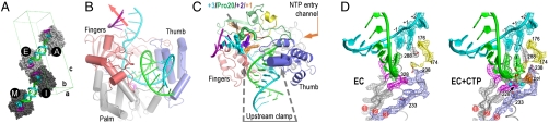
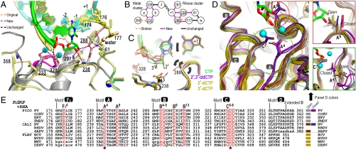
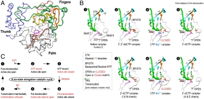
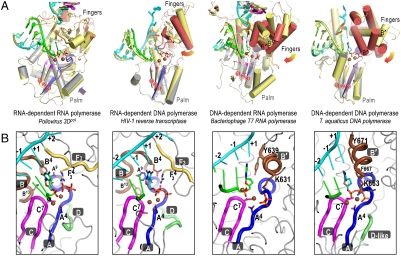
Positive-strand RNA viruses include a large number of human and animal pathogens whose essential RNA-dependent RNA polymerases (RdRPs) share a structurally homologous core with an encircled active site. RdRPs are targets for antiviral drug development, but these efforts are hindered by limited structural information about the RdRP catalytic cycle. To further our understanding of RdRP function, we assembled, purified, and then crystallized poliovirus elongation complexes after multiple rounds of nucleotide incorporation. Here we present structures capturing the active polymerase and its nucleotide triphosphate complexes in four distinct states, leading us to propose a six-state catalytic cycle involving residues that are highly conserved among positive-strand RNA virus RdRPs. The structures indicate that RdRPs use a fully prepositioned templating base for nucleotide recognition and close their active sites for catalysis using a novel structural rearrangement in the palm domain. The data also suggest that translocation by RDRPs may not be directly linked to the conformational changes responsible for active site closure and reopening.


Conflict of interest statement

The authors declare no conflict of interest.

Figures

Fig. 1.
Fig. 1.
Poliovirus 3Dpol elongation complex. (A) Crystal packing showing staggered coaxial stacking of upstream template-product duplexes resulting in two nonequivalent pairs of ECs (A&E vs. I&M). Product strand is shown in green, template in cyan, and downstream nontemplate in purple. (B) EC structure showing up- and downstream RNA duplexes as the template strand (cyan) threads through the active site and the red arrow indicates trajectory of downstream RNA duplex. (C) Top view of EC showing the single stranded conformation of the +1, +2, and +3 downstream template nucleotides and the protein clamp of the upstream duplex. Palm domain is in gray, thumb is in blue, and the individual fingers (19) are colored with index in green, middle in orange, ring in yellow, and the pinky in pink. (D) 3,500 K composite simulated-annealing omit maps contoured at 1.5 σ showing quality of active site electron density for the native (EC) and Mg2+-CTP (EC + CTP) complexes. The pyrophosphate (ppi) in the CTP complex is shown in orange and the presence of metals ions was confirmed by an essentially identical Mn2+-CTP structure (see Figs. S3 and S4).
Fig. 3.
Fig. 3.
Molecular interactions involved in RdRP active site closure. (A) Superposition of 3Dpol active site in open state 1 (yellow) and closed state 4 (colors) showing how ribose hydroxyl recognition drives active site closure and divalent metal binding via the two distinct clusters of interactions diagrammed in panel (B). (C) Comparison of bound CTP, 2′-dCTP, 3′-dCTP, and 2′,3′-ddCTP conformations illustrating the NTP movement associated with active site closure. Only CTP with both ribose hydroxyls triggers the movements (arrows) of Asp238A9 and Ser288B4 that result in the closed conformation and catalysis. β and γ phosphates are omitted for clarity. (D) Conservation of motif A open conformation among all positive-strand RNA virus polymerases shown by superposition of four picornaviral (gray), two caliciviral (olive), and four flaviviral (brown) polymerases and a direct comparison with the closed conformation polio (purple) and Norwalk (blue) virus polymerase structures. Mg2+ ions and newly incorporated CMP are from the state 4 structure and the expanded views show the conformations of key side chains in all the structures. Note that a rabbit hemorrhagic disease virus polymerase structure adopting an intermediate conformation induced by Lu3+ binding is not shown. (E) Structure-based sequence alignment of RdRP motifs A–D and F3. Residues playing key roles in active site interactions and closure are colored red, the two invariant catalytic Asp residues are highlighted by asterisks, and residues in lower case letters either deviate from the consensus structure conformations or are not resolved in the crystal structures and are therefore included based only on sequence homology. See Fig. S8A for an extended alignment of all polymerase motifs across all classes of single-subunit polymerases and Fig. S8B for PDB codes.
Fig. 2.
Fig. 2.
Structures of 3Dpol catalytic cycle states. (A) Superposition of the polymerase structures from all EC states showing small variations in the fingers domain structures and the significant CTP-induced movement of motifs A and D in the palm domain. This movement is highlighted by coloring motif A (red) and motif D (brown) of the closed conformation CTP complexes and showing spheres for the α-carbons of residues 233A4 and 355D5, and the remainder of the structures are colored as in Fig. 1C. (B) Structures of the EC and EC–NTP complexes arranged in order of the proposed catalytic cycle diagrammed in panel C. These include the open conformation state 1 in the absence of NTP, the open state 2 in the presence of bound deoxy-NTPs, the postcatalysis closed conformation state 4 upon CTP addition, and the postcatalysis and pretranslocation open state 5 with 3′-dCTP. Key residues and motifs are indicated in the state 1 panel and the presence of translocation, stacking, and base-pairing interactions involving the templating +1 base and bound nucleotide are indicated. See Figs. S3S7 for electron density maps and additional analyses of the fingers and palm domain movements that take place upon NTP binding.
Fig. 4.
Fig. 4.
(A) A new mode of active site closure in positive-strand RNA virus polymerases. The viral RdRPs close their active site using a rearrangement of the palm domain that results in the complete formation of the 3-stranded palm domain β-sheet common to all polymerases. In contrast, representative structures from the other three classes of polymerases show major swinging motions of their fingers domains between the open (fingers yellow, motif A brown) and closed (fingers red, motif A blue) conformations that serve to reposition the +1 templating base (orange) and incoming nucleotide for catalysis. For clarity, only the closed conformation NTP, 6-bp of upstream duplex, and part of the protein structures are shown and spheres correspond to catalytic metals and the motif A AspA4 Cα atom. The motif B’ helix that plays a major role in translocation by the DNA-templated polymerases is labeled at its N-terminal end. (B) The common organization of the closed conformation active sites across all four classes of polymerases that is built on the 3-stranded antiparallel β-sheet of motifs A and C in the palm domain. In comparing the RNA- and DNA-templated polymerases, note the structural divergence of the B and B’ motifs and how the F3 and B’ motifs provides analogous charge interactions with the bound nucleotide. Conserved motifs A–D are indicated and key residues are labeled according to the alignment shown in Fig. 3E. See Figs. S6 and S8 for additional details.

References

    1. Ng KK, Arnold JJ, Cameron CE. Structure-function relationships among RNA-dependent RNA polymerases. Curr Top Microbiol Immunol. 2008;320:137–156. - PMC - PubMed
    1. Ferrer-Orta C, Arias A, Escarmis C, Verdaguer N. A comparison of viral RNA-dependent RNA polymerases. Curr Opin Struct Biol. 2006;16:27–34. - PubMed
    1. Lescar J, Canard B. RNA-dependent RNA polymerases from flaviviruses and Picornaviridae. Curr Opin Struct Biol. 2009;19:759–767. - PubMed
    1. Bruenn JA. A structural and primary sequence comparison of the viral RNA-dependent RNA polymerases. Nucleic Acids Res. 2003;31:1821–1829. - PMC - PubMed
    1. Poch O, Sauvaget I, Delarue M, Tordo N. Identification of four conserved motifs among the RNA-dependent polymerase encoding elements. EMBO J. 1989;8:3867–3874. - PMC - PubMed

Publication types

MeSH terms