. 2011 Mar;300(3):L362-9.
doi: 10.1152/ajplung.00278.2010. Epub 2010 Dec 10.

Mitigation of chlorine gas lung injury in rats by postexposure administration of sodium nitrite

Affiliations

Mitigation of chlorine gas lung injury in rats by postexposure administration of sodium nitrite

Amit K Yadav et al. Am J Physiol Lung Cell Mol Physiol. 2011 Mar.

Abstract

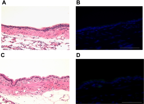
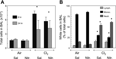
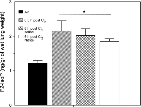
Nitrite (NO(2)(-)) has been shown to limit injury to the heart, liver, and kidneys in various models of ischemia-reperfusion injury. Potential protective effects of systemic NO(2)(-) in limiting lung injury or enhancing repair have not been documented. We assessed the efficacy and mechanisms by which postexposure intraperitoneal injections of NO(2)(-) mitigate chlorine (Cl(2))-induced lung injury in rats. Rats were exposed to Cl(2) (400 ppm) for 30 min and returned to room air. NO(2)(-) (1 mg/kg) or saline was administered intraperitoneally at 10 min and 2, 4, and 6 h after exposure. Rats were killed at 6 or 24 h. Injury to airway and alveolar epithelia was assessed by quantitative morphology, protein concentrations, number of cells in bronchoalveolar lavage (BAL), and wet-to-dry lung weight ratio. Lipid peroxidation was assessed by measurement of lung F(2)-isoprostanes. Rats developed severe, but transient, hypoxemia. A significant increase of protein concentration, neutrophil numbers, airway epithelia in the BAL, and lung wet-to-dry weight ratio was evident at 6 h after Cl(2) exposure. Quantitative morphology revealed extensive lung injury in the upper airways. Airway epithelial cells stained positive for terminal deoxynucleotidyl-mediated dUTP nick end labeling (TUNEL), but not caspase-3. Administration of NO(2)(-) resulted in lower BAL protein levels, significant reduction in the intensity of the TUNEL-positive cells, and normal lung wet-to-dry weight ratios. F(2)-isoprostane levels increased at 6 and 24 h after Cl(2) exposure in NO(2)(-)- and saline-injected rats. This is the first demonstration that systemic NO(2)(-) administration mitigates airway and epithelial injury.


Figures

Fig. 1.
Fig. 1.
Breathing frequencies and peripheral oxygen saturations. Rats were exposed to 400 ppm Cl2 for 30 min, returned to room air, and killed 6 h (A and C) or 24 h (B and D) after exposure. All rats received intraperitoneal injections of NO2 (1 mg/kg body wt) or an equivalent amount of saline (50 μl) at 0.1, 2, 4, and 6 h after exposure. Values are means ± SE. A and B: breath rates (determined by observation). C and D: peripheral oxygen saturations. In A and C, n = 8 air + saline and air + NO2, n = 7 Cl2 + saline, n = 9 Cl2 + NO2; in B and D, n = 3 air + saline and air + NO2, n = 6 Cl2 + saline and Cl2 + NO2. Breathing frequencies and oxygen saturations for Cl2 + saline and Cl2 + NO2 at all time points, except 24 h after exposure, are significantly different from their corresponding control values (1-way ANOVA followed by Tukey-Kramer multiple-comparisons test).
Fig. 2.
Fig. 2.
NO2 decreases protein levels in bronchoalveolar lavage (BAL) and bloodless lung wet-to-dry weight ratios. Rats were exposed to 400 ppm Cl2 for 30 min, returned to room air, and killed 6 or 24 h after exposure. All rats received intraperitoneal injections of NO2 (1 mg/kg body wt) or an equivalent amount of saline (200–250 μl) at 0.1, 2, 4, and 6 h after exposure. At 6 or 24 h after exposure, they were killed and their lungs were lavaged with 8 ml of normal saline, which was instilled and withdrawn slowly 3 times; BAL samples were spun at 300 g for 10 min at 4°C to pellet cells (22). A: protein concentrations in cell-free BAL measured using the bicinchoninic acid (BCA) Protein Assay Reagent Kit (Pierce, Rockford, IL) and as previously described (17). B: blood-free lung wet-to-dry weight (W/D) ratios. Values are means ± SE; n = 8 air + saline, n = 2 air + NO2, n = 7 Cl2 + saline, n = 10 Cl2 + NO2 at 6 h; n = 3 air + NO2, n = 6 Cl2 + saline and Cl2 + NO2 at 24 h; for wet-to-dry lung weights, n = 4 for each group. *P < 0.05 vs. corresponding air value (ANOVA followed by Tukey's t-test for multiple comparisons). #P < 0.05 vs. corresponding saline value (2-tailed t-test). ^P < 0.05 vs. corresponding saline value (1-tailed t-test).
Fig. 3.
Fig. 3.
Proximal airways of rats exposed to air and injected with NO2. A and C: hematoxylin-and-eosin-stained sections of rat proximal airways injected with saline (A) or NO2 (C). Note normal lung architecture with prominent cilia. B and D: adjacent sections processed for terminal deoxynucleotidyl-mediated dUTP nick end labeling (TUNEL) using the DeadEnd Fluorometric TUNEL System and imaged with a fluorescent microscope for detection of FITC (green color). Note absence of green fluorescence, indicating absence of apoptosis/necrosis. Approximately 5 sections were examined from each rat (n = 3) with similar results. Scale bar, 50 μm.
Fig. 4.
Fig. 4.
Exposure to Cl2 causes extensive injury to proximal lung airway epithelia. Rats were exposed to Cl2 (400 ppm for 30 min), returned to room air, injected with saline (A and B) or NO2 (C and D), and killed 6 h after exposure. A and C: hematoxylin-and-eosin-stained sections of proximal airway. Note sloughed airway epithelium and injury to the interstitial space (A; saline) and relatively less sloughing of airway epithelium (C; NO2). B and D: adjacent sections processed for TUNEL using the DeadEnd Fluorometric TUNEL system and imaged with a fluorescence microscope for detection of FITC (green color) and 4′,6-diamidino-2-phenylindole (blue fluorescence) to identify nuclei. Sloughed portion of the epithelium in A stained positive, as indicated by considerable green fluorescence (B). Very little green fluorescence is seen in D. Approximately 5 sections were examined from each rat (n = 3) with similar results. Scale bar, 50 μm.
Fig. 5.
Fig. 5.
Quantification of TUNEL in lung airways of saline- and NO2-injected animals exposed to Cl2. Rats were exposed to Cl2 (400 ppm for 30 min), returned to room air, and killed 6 h after exposure. They received injections of NO2 or saline. Average intensity of green fluorescence levels in main bronchus images (see Fig. 4) was calculated using Metamorph imaging software by 2 different investigators who were blinded regarding the groups of the rats. Five images were obtained for each rat, which were averaged so that each rat was weighted equally. Values are means ± SE; n = 3 rats in each group. *P < 0.001 vs. corresponding control value. #P < 0.001 vs. corresponding saline value in the same group.
Fig. 6.
Fig. 6.
Airway sections of rats exposed to Cl2 and killed 24 h after exposure. Rats were exposed to 400 ppm Cl2 for 30 min, returned to room air, injected with saline (A and B) or NO2 (C and D), and killed 24 h after exposure. A and C: hematoxylin-and-eosin-stained sections of proximal airways. Note absence of epithelium in saline-injected rat (A) and presence of pseudostratified epithelium, indicating onset of repair, in NO2-injected rat. Only background levels of green fluorescence were seen in the adjoining sections (B and D) processed for TUNEL, indicating the absence of necrotic/apoptotic cells. Approximately 5 sections were examined from each rat (n = 3) with similar results. Scale bar, 50 μm.
Fig. 7.
Fig. 7.
Injections of NO2 in Cl2-exposed rats do not decrease the number of inflammatory cells in BAL. Rats were exposed to 400 ppm Cl2 for 30 min, returned to room air, and killed 6 or 24 h after exposure. All rats received intraperitoneal injections of NO2 (1 mg/kg body wt in 50 μl of normal saline) or an equivalent amount of saline at 0.1, 2, 4, and 6 h after exposure. Values are means ± SE; n = 8 animals in 6-h group and 3 animals in 24-h group. *P < 0.05 vs. corresponding air values.
Fig. 8.
Fig. 8.
Exposure to Cl2 increases F2-isoprostane (F2-IsoP) levels in lung tissues. Rats were exposed to 400 ppm Cl2 for 30 min, returned to room air, and killed 6 h after exposure. All rats received intraperitoneal injections of NO2 (1 mg/kg body wt in 50 μl of normal saline) or an equivalent amount of saline at 0.1, 2, 4, and 6 h after exposure. Values are means ± SE; n = 8 in each group. *P < 0.05 vs. corresponding air values.

References

    1. Andonegui G, Goyert SM, Kubes P. Lipopolysaccharide-induced leukocyte-endothelial cell interactions: a role for CD14 versus toll-like receptor 4 within microvessels. J Immunol 169: 2111–2119, 2002 - PubMed
    1. Baskin SI, Horowitz AM, Nealley EW. The antidotal action of sodium nitrite and sodium thiosulfate against cyanide poisoning. J Clin Pharmacol 32: 368–375, 1992 - PubMed
    1. Bell DG. Management of acute respiratory distress syndrome (ARDS) following chlorine exposure (Abstract). Am J Respir Crit Care Med 176: A314, 2008
    1. Comellas AP, Pesce LM, Azzam Z, Saldias FJ, Sznajder JI. Scorpion venom decreases lung liquid clearance in rats. Am J Respir Crit Care Med 167: 1064–1067, 2003 - PubMed
    1. Crawford JH, Isbell TS, Huang Z, Shiva S, Chacko BK, Schechter AN, Darley-Usmar VM, Kerby JD, Lang JD, Jr, Kraus D, Ho C, Gladwin MT, Patel RP. Hypoxia, red blood cells, and nitrite regulate NO-dependent hypoxic vasodilation. Blood 107: 566–574, 2006 - PMC - PubMed

Publication types

MeSH terms