. 2011 Jan 15;186(2):1189-98.
doi: 10.4049/jimmunol.1001794. Epub 2010 Dec 10.

The human glucocorticoid receptor as an RNA-binding protein: global analysis of glucocorticoid receptor-associated transcripts and identification of a target RNA motif

Affiliations

The human glucocorticoid receptor as an RNA-binding protein: global analysis of glucocorticoid receptor-associated transcripts and identification of a target RNA motif

Faoud T Ishmael et al. J Immunol. .

Abstract

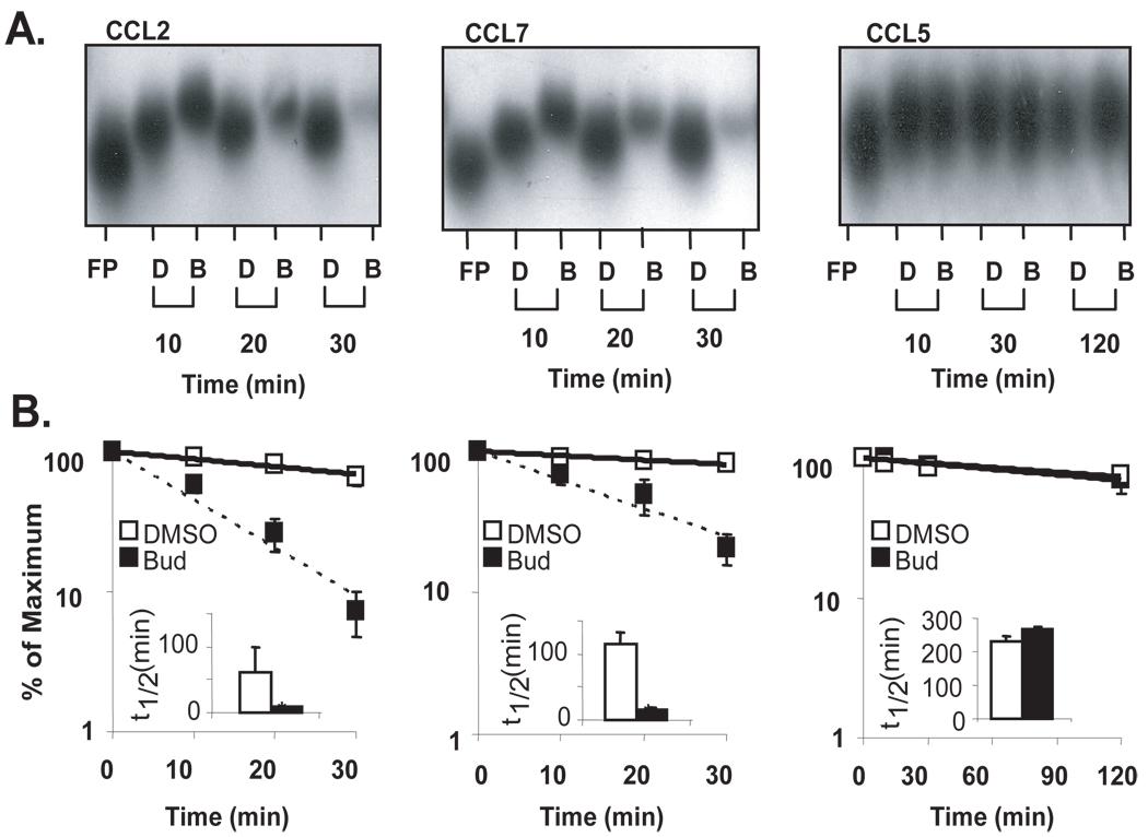
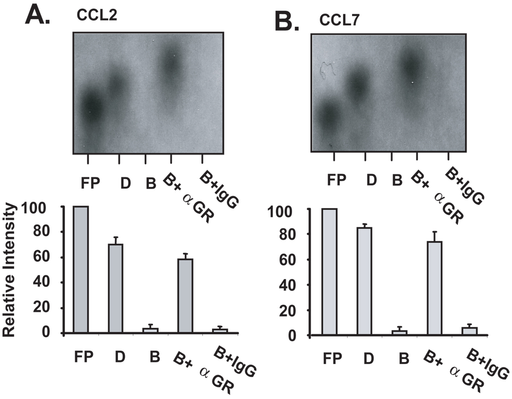
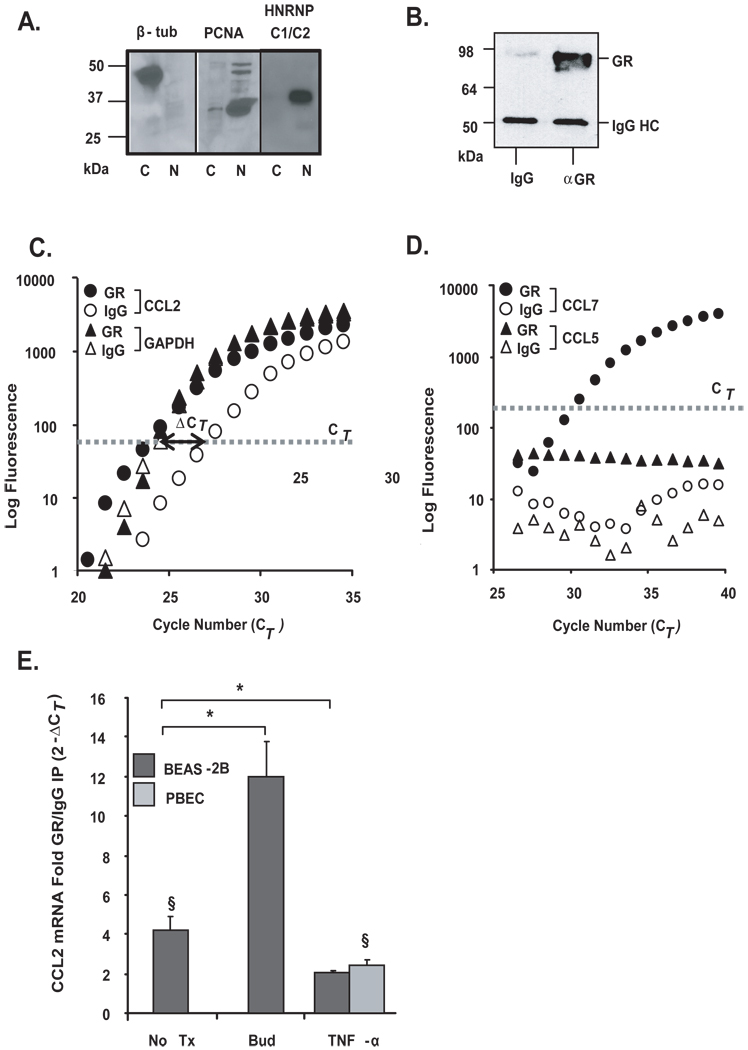
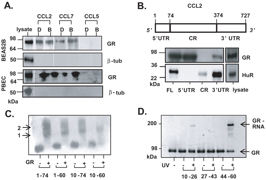
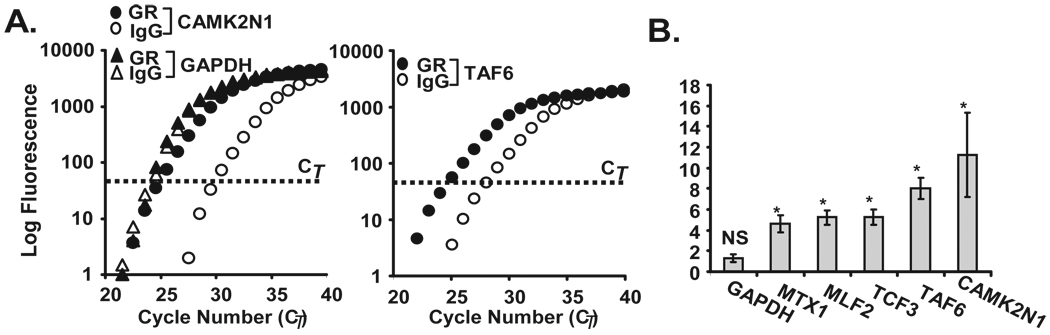
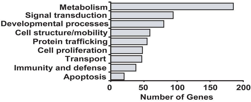
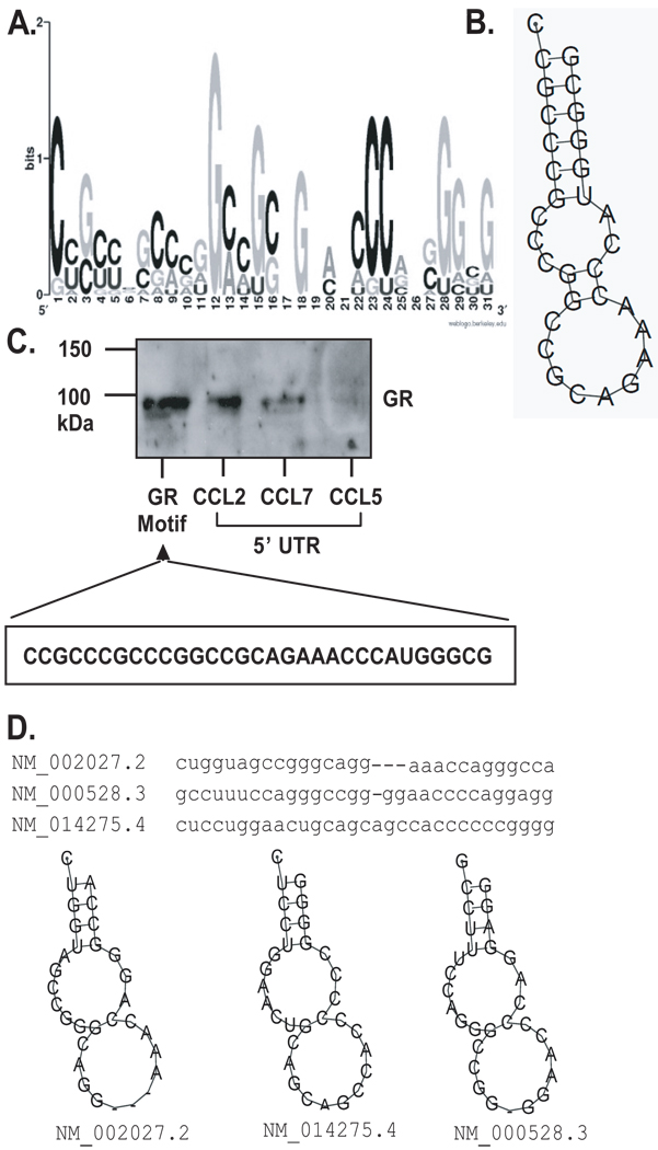
Posttranscriptional regulation is emerging as a key factor in glucocorticoid (GC)-mediated gene regulation. We investigated the role of the human GC receptor (GR) as an RNA-binding protein and its effect on mRNA turnover in human airway epithelial cells. Cell treatment with the potent GC budesonide accelerated the decay of CCL2 mRNA (t(1/2) = 8 ± 1 min versus 62 ± 17 min in DMSO-treated cells) and CCL7 mRNA (t(1/2) = 15 ± 4 min versus 114 ± 37 min), but not that of CCL5 mRNA (t(1/2)=231 ± 8 min versus 266 ± 5 min) in the BEAS-2B cell line. This effect was inhibited by preincubation with an anti-GR Ab, indicating that GR itself plays a role in the turnover of these transcripts. Coimmunoprecipitation and biotin pulldown experiments showed that GR associates with CCL2 and CCL7 mRNAs, but not CCL5 mRNA. These methods confirmed CCL2 mRNA targeting by GR in human primary airway epithelial cells. Association of the GR was localized to the 5' untranslated region of CCL2 mRNA and further mapped to nt 44-60. The collection of transcripts associated with GR, identified by immunoprecipitation of GR-mRNA complexes followed by microarray analysis, revealed 479 transcripts that associated with GR. Computational analysis of the primary sequence and secondary structures of these transcripts yielded a GC-rich motif, which was shown to bind to GR in vitro. This motif was used to predict binding of GR to an additional 7889 transcripts. These results indicate that cytoplasmic GR interacts with a subset of mRNA through specific sequences and can regulate turnover rates, suggesting a novel posttranscriptional role for GR as an RNA-binding protein.


Figures

Figure 1
Figure 1. Glucocorticoids promote selective degradation of CCL2 and CCL7 mRNAs in a cell-free assay of mRNA decay
A. Non-denaturing PAGE (representative of n=3) of in vitro transcribed, biotinylated human CCL2, CCL7, or CCL5 full-length mRNAs alone [lane 1, Free Probe (FP)] or mixed with cytoplasmic lysates of BEAS-2B cells (pre-treated with budesonide (B, 10−7 M) or DMSO (D) for 3h) for the indicated times. B. Densitometric measurement of the amount of mRNA at each time point; mean ± SEM, n=3. Inset bargraphs: Half-lives of chemokine mRNA in each condition (calculated from plots in B, as Ln(0.5)/slope, indicating the time at which 50% of the initial mRNA is left); * p<0.05.
Figure 2
Figure 2. Treatment of BEAS-2B cell lysates with a specific antibody against hGR inhibits GC-mediated decay of CCL2 and CCL7 mRNAs
Non-denaturing PAGE (representative of n=3) of biotinylated human CCL2 (A) or CCL7 (B) mRNA incubated with cytoplasmic lysates (2 µg) of DMSO-(D) or budesonide-treated (B) BEAS-2B cells. Budesonide-treated lysates were pre-incubated with 3 µg of an anti-GR antibody (B+αGR) or isotype control antibody (B+IgG) for 30 minutes before addition of labeled RNA. Samples were further incubated for 30 minutes and then subjected to PAGE. The relative intensity of each band compared to the free probe was determined by densitometry (shown in bar graphs below is densitometric mean ± SEM of n=3 for each chemokine).
Figure 3
Figure 3. Association of chemokine transcripts with GR in airway epithelial cells
A. The purity of cytoplasmic lysates was verified by Western blot analysis of a cytoplasmic protein [β-tubulin (β-tub)] and nuclear proteins (PCNA, HNRNP C1/C2), detected exclusively in cytoplasmic (c) and nuclear (n) extracts, respectively. B. Western Blot analysis of GR after immunoprecipitation (IP) of protein-mRNA complexes obtained using the monoclonal anti-GR antibody (αGR) or the IgG-control antibody control (IgG), showing selectivity of the IP. Bands corresponding to GR and IgG heavy chain (IgG HC) are indicated. C. Real-time PCR amplification plot of CCL2 and GAPDH mRNA (representative of n=10) and D. of CCL7 and CCL5 mRNA (representative of n=5), both after GR (filled circles) or IgG control (open circles) IP. The bold line with arrow points indicates the difference in Ct (ΔCT) between the CCL2 mRNA detected in the GR-IP versus the IgG-IP. E. Mean ± SEM fold GR/IgG IP enrichment for CCL2 mRNA (expressed as 2−ΔCT) in BEAS-2B and PBEC cells unstimulated (n=10) or treated with budesonide (bud, n=4) or TNFα (n=3). * p < 0.03 for treated samples vs. untreated, § p < 0.03 vs. GR-IP control.
Figure 4
Figure 4. Localization of GR binding on the 5’UTR of CCL2 mRNA
A. Western blot analysis of biotin pull-down assay showing association of GR from cytoplasmic extracts of BEAS-2B (n=3) and PBEC (n=2) treated with budesonide (B, 10−7 M) or DMSO (D), incubated with biotinylated transcripts spanning human CCL2 or CCL7 full-length mRNA. In both cell types, lack of β-tubulin detection was used to rule out non-specific binding. B. Above: schematic of CCL2 mRNA. Indicated are the nucleotide numbers corresponding to untranslated regions (UTR) and coding region (CR). Below, detection of GR by Western blot after biotin pulldown assay from unstimulated BEAS-2B cell lysate, using biotinylated transcripts encompassing the CCL2 full-length RNA (FL), the 5’ and 3’ UTRs, or the CR. The RBP HuR is detected as positive control for association with CCL2 3’UTR. C. EMSA of purified, recombinant GR incubated with mRNA probes encompassing discrete regions of CCL2 5’UTR (indicated by nucleotide number). Arrows represent putative GR monomer (1) and dimer (2) binding. C. UV cross-linking of purified GR protein to 15-nt fragments of the CCL2 5’UTR, followed by detection by Western blot of the covalently-linked GR via mobility shift of the RNA-bound GR protein (GR-RNA).
Figure 5
Figure 5. Validation of GR-associated targets identified by gene array
A. Real-time PCR amplification plot (representative of n=3) after GR (filled circles) or control (open circles) IP for CAMK2N1and TAF6, identified as GR-associated transcripts by the GR-IP array study, and GAPDH as control. B. Bargraph represent the mean ± SEM fold GR/IgG IP enrichment for MTX1, MLF2, TCF3, TAF6, and CAMK2N1 (n=3; *, p<0.05 compared to IgG IP, NS=not significant for GAPDH).
Figure 6
Figure 6. Genome Ontology Analysis of GR-associated genes
The analysis was performed on the full list of 477 putative GR targets (listed in full in the Supplemental Table2) identified by hybridization of unstimulated BEAS-2B cDNAs to an Illumina human cDNA array using GR-IP arrays versus IgG control-IP arrays.
Figure 7
Figure 7. Primary sequence and secondary structure of the predicted GR mRNA motif
A. Graphic logo representing the probability matrix of the GR motif, showing the relative frequency of each nucleotide for each position within the motif sequence. The motif is originated from the experimental data set from the array study. B. Secondary structure of the GR motif comprising the nucleotides with highest frequency for each position within the motif shown in A. C. Biotin pull-down assay showing association of GR from unstimulated BEAS-2B cell lysates with the GR motif shown in B., compared to GR association with the full-length 5’UTR of CCL2, CCL7 and CCL5 mRNA (the latter as negative control). D. Sequence and secondary structure of the GR motif in three mRNAs from the UniGene list of putative GR targets; the corresponding RefSeq accession numbers and names are shown.

References

    1. Barnes PJ. How corticosteroids control inflammation: Quintiles Prize Lecture 2005. Br J Pharmacol. 2006;148:245–254. - PMC - PubMed
    1. Stellato C. Glucocorticoid actions on airway epithelial responses in immunity: functional outcomes and molecular targets. J Allergy Clin Immunol. 2007;120:1247–1263. quiz 1264-1245. - PubMed
    1. Adcock IM, Ito K, Barnes PJ. Glucocorticoids: Effects on Gene Transcription. Proc Am Thorac Soc. 2004;1:247–254. - PubMed
    1. Bhavsar P, Hew M, Khorasani N, Torrego A, Barnes PJ, Adcock I, Chung KF. Relative corticosteroid insensitivity of alveolar macrophages in severe asthma compared with non-severe asthma. Thorax. 2008;63:784–790. - PubMed
    1. Rhen T, Cidlowski JA. Antiinflammatory Action of Glucocorticoids -- New Mechanisms for Old Drugs. N Engl J Med. 2005;353:1711–1723. - PubMed

Publication types

MeSH terms

Associated data