. 2011 Apr;39(8):3363-72.
doi: 10.1093/nar/gkq946. Epub 2010 Dec 11.

Mechanistic insights into an engineered riboswitch: a switching element which confers riboswitch activity

Affiliations

Mechanistic insights into an engineered riboswitch: a switching element which confers riboswitch activity

Julia E Weigand et al. Nucleic Acids Res. 2011 Apr.

Abstract

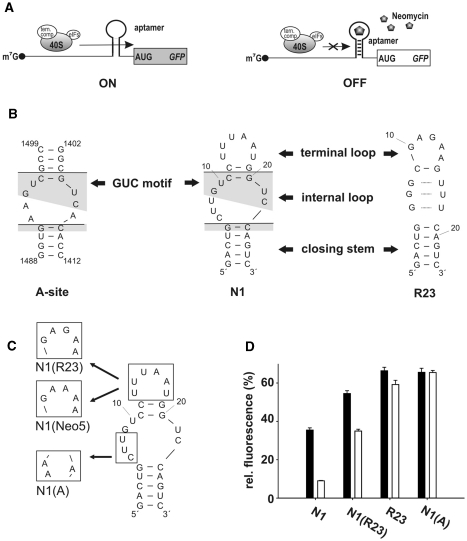
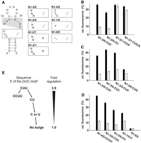
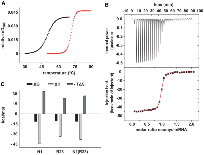
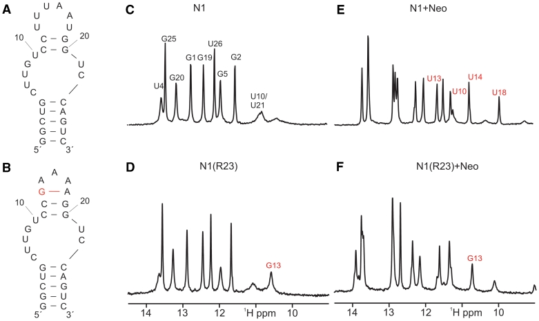
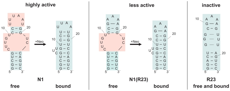
While many different RNA aptamers have been identified that bind to a plethora of small molecules only very few are capable of acting as engineered riboswitches. Even for aptamers binding the same ligand large differences in their regulatory potential were observed. We address here the molecular basis for these differences by using a set of unrelated neomycin-binding aptamers. UV melting analyses showed that regulating aptamers are thermally stabilized to a significantly higher degree upon ligand binding than inactive ones. Regulating aptamers show high ligand-binding affinity in the low nanomolar range which is necessary but not sufficient for regulation. NMR data showed that a destabilized, open ground state accompanied by extensive structural changes upon ligand binding is important for regulation. In contrast, inactive aptamers are already pre-formed in the absence of the ligand. By a combination of genetic, biochemical and structural analyses, we identified a switching element responsible for destabilizing the ligand free state without compromising the bound form. Our results explain for the first time the molecular mechanism of an engineered riboswitch.


Figures

Figure 1.
Figure 1.
Conditional control by neomycin-binding aptamers. (A) Scheme of aptamer-mediated inhibition of translation initiation. The aptamer is inserted directly upstream of the start codon. Left: without ligand the aptamer does not interfere with ribosomal scanning. Right: the aptamer ligand complex inhibits the small ribosomal subunit and leads to decreased gene expression. (B) Secondary structure of the ribosomal A-site and the neomycin-binding aptamers N1 and R23. The lines dissect the aptamers into the terminal loop, an internal asymmetrical loop and the closing stem. Conserved nucleotides between the A-site and N1 are shaded in gray. (C) Hybrid aptamers. (D) gfp expression in the absence (black bars) and presence (white bars) of 100 µM neomycin. The fluorescence emission of the vector pWHE601 (6) expressing gfp without an aptamer in its 5′UTR was set as 100%. Background level of a vector with no gfp expression was subtracted from all data. Values represent the mean of three independently grown cultures. Measurements were repeated at least twice.
Figure 2.
Figure 2.
Saturating mutagenesis of the internal loop. (A) Secondary structure of N1. The GUC motif is shaded in gray. The lower part of the internal loop (boxed) was analyzed and randomized. Nucleotides are indicated with N (any nucleotide) or Y (pyrimidines). (B–D) gfp expression in the absence (black bars) and presence (white bars) of 100 µM neomycin. The fluorescence emission of the vector pWHE601 (6) expressing gfp without an aptamer in its 5′UTR was set as 100%. Background level of a vector with no gfp expression was subtracted from all data. Values represent the mean of three independently grown cultures. Measurements were repeated at least twice. (E) Schematic representation of the connection between loop size and regulation.
Figure 3.
Figure 3.
Melting point analyses and determination of the equilibrium dissociation constants (KD) by ITC. (A) Melting curves in the absence (black curves) and presence (red curves) of 10 µM neomycin for 1 µM N1. Melting curves were recorded in triplicates. (B) Upper panel: power required to maintain the temperature (37°C) of the RNA solution (4 µM) recorded over the time of multiple injections (10 µl) of ligand (38 µM neomycin) until saturation was reached (baseline-corrected). Lower panels: integrated heats of interaction per mole of injectant plotted against the molar ratio of ligand over RNA and fitted to a single binding site model. (C) Gibbs free energies (ΔG), enthalpic (ΔH) and entropic (–TΔS) contributions of N1, R23 and N1(R23).
Figure 4.
Figure 4.
Conformation and neomycin binding of N1 and N1(R23). (A and B) Proposed secondary structure of the free form of N1 and N1(R23) based on the number of observable imino proton signals and NOE-patterns. The additional base pair in the terminal loop of N1(R23) is highlighted in red. (C and D) Comparison of the imino proton region of 1D-1H-spectra of free N1 and free N1(R23). Signal assignments are indicated. The assignment of the signal at ∼10.7 ppm to the imino proton of G13 (marked in red) is based on the comparison with spectra of the original R23 aptamer in its ligand-free state (20). (E and F) Imino proton region of the 1D-1H-spectrum of N1 and N1(R23) in the presence of one equivalent of neomycin. Compared to the spectrum of free N1 and N1(R23) shown in (D and E) novel signals and chemical shift changes are observed as expected due to the formation of stable 1:1 neomycin RNA-complexes in slow exchange on the NMR-time scale.
Figure 5.
Figure 5.
Model of proposed secondary structure changes of N1, N1(R23) and R23 upon neomycin binding derived from NMR data. Unstructured elements are shaded in red, structured elements in blue.

References

    1. Roth A, Breaker RR. The structural and functional diversity of metabolite-binding riboswitches. Annu. Rev. Biochem. 2009;78:305–334. - PMC - PubMed
    1. Suess B, Weigand JE. Engineered riboswitches: overview, problems and trends. RNA Biol. 2008;5:24–29. - PubMed
    1. Weigand JE, Suess B. Aptamers and riboswitches: perspectives in biotechnology. Appl. Microbiol. Biotechnol. 2009;85:229–236. - PubMed
    1. Hermann T, Patel DJ. Adaptive recognition by nucleic acid aptamers. Science. 2000;287:820–825. - PubMed
    1. Hanson S, Berthelot K, Fink B, McCarthy JE, Suess B. Tetracycline-aptamer-mediated translational regulation in yeast. Mol. Microbiol. 2003;49:1627–1637. - PubMed

Publication types