. 2010 Dec 13;191(6):1159-72.
doi: 10.1083/jcb.201005060.

Formin follows function: a muscle-specific isoform of FHOD3 is regulated by CK2 phosphorylation and promotes myofibril maintenance

Affiliations

Formin follows function: a muscle-specific isoform of FHOD3 is regulated by CK2 phosphorylation and promotes myofibril maintenance

Thomas Iskratsch et al. J Cell Biol. .

Abstract

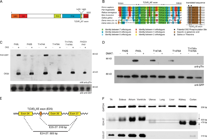
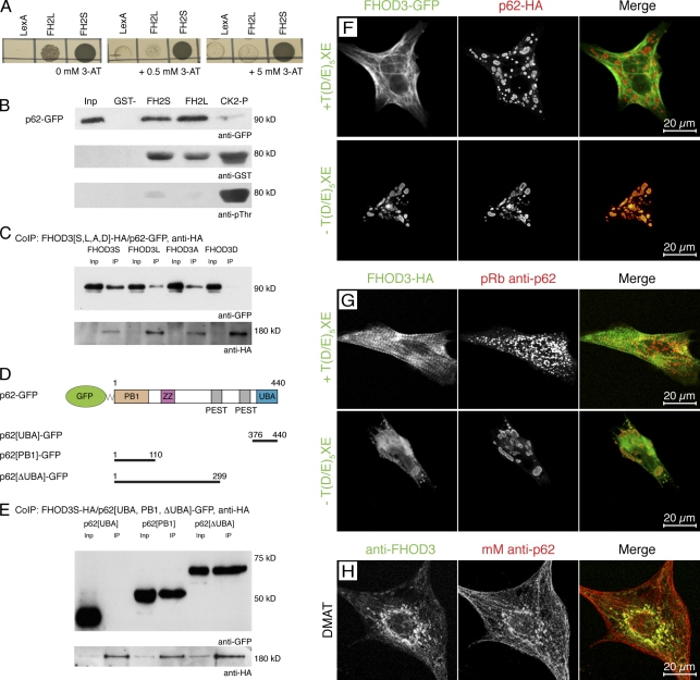
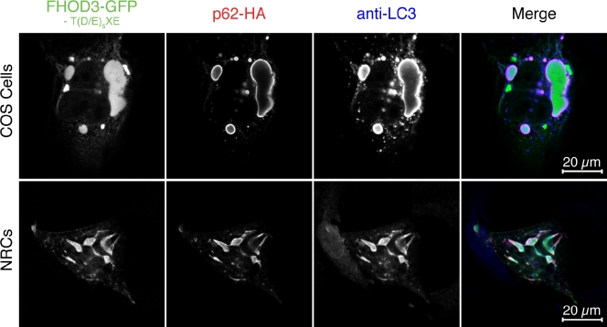
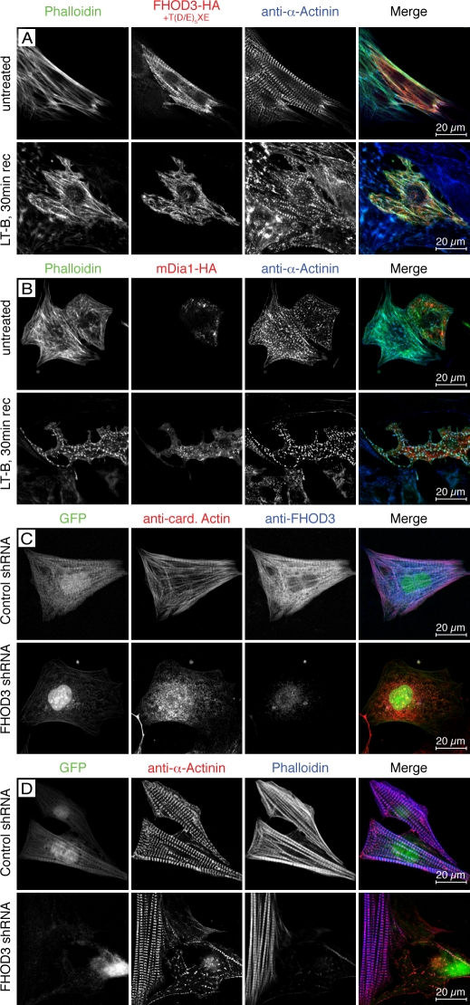
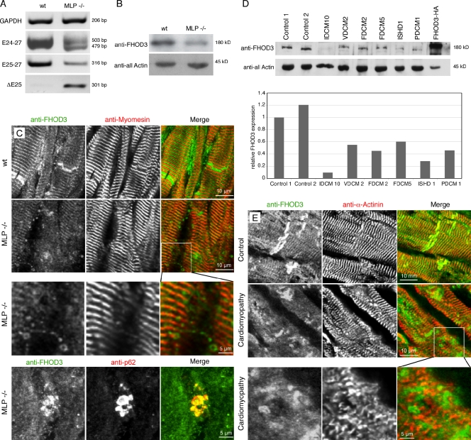
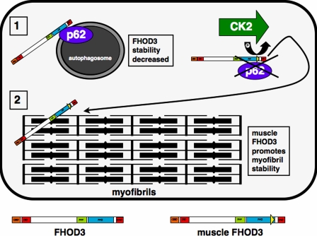
Members of the formin family are important for actin filament nucleation and elongation. We have identified a novel striated muscle-specific splice variant of the formin FHOD3 that introduces a casein kinase 2 (CK2) phosphorylation site. The specific targeting of muscle FHOD3 to the myofibrils in cardiomyocytes is abolished in phosphomutants or by the inhibition of CK2. Phosphorylation of muscle FHOD3 also prevents its interaction with p62/sequestosome 1 and its recruitment to autophagosomes. Furthermore, we show that muscle FHOD3 efficiently promotes the polymerization of actin filaments in cardiomyocytes and that the down-regulation of its expression severely affects myofibril integrity. In murine and human cardiomyopathy, we observe reduced FHOD3 expression with a concomitant isoform switch and change of subcellular targeting. Collectively, our data suggest that a muscle-specific isoform of FHOD3 is required for the maintenance of the contractile structures in heart muscle and that its function is regulated by posttranslational modification.


Figures

Figure 1.
Figure 1.
FHOD3 contains a novel alternative exon at the end of its FH2 domain. (A) Domain layout of the human FHOD3 splice isoform expressing the T(D/E)5XE exon (exon 26 in humans and exon 25 in rodents; Fig. S1). GBD, GTPase-binding domain; DID, diaphanous inhibitory domain; FH1/FH2, formin homology domains 1 and 2. (B) The alternative T(D/E)5XE exon of FHOD3 encodes for an evolutionarily conserved stretch of eight mainly acidic amino acids that introduce CK2 consensus sites. Lowercase letters are intronic, and capital letters represent exonic sequences. Splice acceptor and donor sites are evolutionarily conserved in all seven orthologues, with the intronic bases AG on the 5′ end and GT on the 3′ end. (C) GST fusion proteins of the FHOD3-FH2 domain containing (FH2L) or excluding (FH2S) the alternative T(D/E)5XE exon, as well as threonine to alanine mutants of the predicted target threonines (T1474, T1476, or both), and the FHOD1-FH2 domain were tested for CK2 in vitro phosphorylation. FH2L, but not FH2S or FHOD1-FH2, was phosphorylated by CK2. Mutation of T1474 or T1476 to alanine reduced phosphorylation slightly or greatly, whereas mutation of both residues completely abolished the phosphorylation. mM, monoclonal mouse. (D) A similar phosphorylation pattern to that seen in vitro could also be demonstrated in COS-1 cells transfected with different GFP-tagged FHOD3 constructs and was abolished by treatment with 10 µM of the CK2 inhibitor DMAT for 24 h before lysis. The signal for GFP is shown as a loading control below. (E) Schematic view of the primer sets used for the RT-PCRs. (F) RT-PCR to test for tissue-specific expression of the alternative T(D/E)5XE exon. The amount of template mouse cDNAs was normalized against glycerine aldehyde 3-phosphate dehydrogenase (GAPDH). The specificity of the T(D/E)5XE exon for striated muscle can either be seen with primer set E25-27 or, because of the difference in the electrophoretic mobility, with primer set E24-27. The top line points out the 503-bp fragment, and the bottom line indicates the 479-bp fragment, which are also indicated by the arrows. TA, tibialis anterior.
Figure 2.
Figure 2.
The muscle-specific FHOD3 isoform is targeted to the sarcomere in a phosphorylation-dependent fashion. (A and B) NRCs were transfected with FHOD3-HA including or lacking the alternative T(D/E)5XE exon (A) or after mutation of residues T1474 and T1476 to alanine (T>A) or aspartate (T>D; B). After 48 h, cells were fixed and stained with monoclonal rat anti-HA and pRb anti–myosin-binding protein C (MyBP-C). Constructs including the T(D/E)5XE exon targeted to the A band, whereas constructs lacking the T(D/E)5XE exon formed cytoplasmic aggregates. Mutation of the threonine residues to alanine induced the formation of cytoplasmic aggregates as well. No change in localization was seen after mutation of the threonine residues to aspartic acid. (C) Untransfected NRCs were treated with 10 µM of the CK2 inhibitor DMAT for 24 h and then fixed and stained with anti-FHOD3 and anti–α-actinin antibodies. Untreated cells were used as a control. Similar to exogenous FHOD3, endogenous FHOD3 localizes to the A band but forms aggregates when phosphorylation by CK2 is inhibited.
Figure 3.
Figure 3.
FHOD3 interacts with the p62-PB1 domain in a phosphorylation-regulated manner. (A) Yeast two-hybrid assay to test for the specificity and strength of the p62 interaction with the FHOD3-FH2 domain. p62-pGAD10 was transformed into yeast together with FH2L and FH2S-LexA, as well as with the empty LexA vector as a control, and grown on medium containing 0, 0.5, and 5.0 mM of 3-AT. p62 strongly interacts with FH2S and interacts much more weakly with FH2L. (B) GST pull-down assays to analyze the FHOD3–p62 interaction. Equal amounts of purified FH2L-GST, FH2S-GST, or CK2-phosphorylated FH2L-GST (CK2-P) were incubated with lysates of p62-GFP–transfected COS-1 cells and subjected to immunoblotting with monoclonal mouse (mM) anti-GFP and, subsequently, polyclonal goat anti-GST. The successful phosphorylation of FH2L-GST was controlled by incubation with monoclonal mouse anti–phospho-threonine (pThr). Although the presence of the unphosphorylated T(D/E)5XE exon has no effect on the interaction with p62, CK2 phosphorylation of FH2L-GST before the incubation strongly reduces the interaction. Inp, input. (C) Coimmunoprecipitation (IP; CoIP) of full-length FHOD3 and p62. Lysates of COS-1 cells, which were transfected with p62-GFP and FHOD3-HA constructs containing (FHOD3L) or excluding (FHOD3S) the T(D/E)5XE exon, as well as with constructs with residues T1474 and T1476 mutated to alanine (FHOD3A) or aspartic acid (FHOD3D), were immunoprecipitated with anti-HA and subjected to immunoblotting with anti-GFP and subsequently anti-HA antibodies. Mutation of the threonine residues to aspartic acid, but not to alanine, greatly reduced the interaction. (D) p62 domain structure and transfection constructs. Domains were constructed according to Simple Modular Architecture Research Tool (SMART) and Geetha and Wooten (2002). ZZ, zinc finger domain (ZZ type). (E) To identify the minimal binding site, COS-1 cells were cotransfected with FHOD3S-HA and p62-GFP constructs containing the PB1 domain, the UBA domain, or the protein without the UBA domain (ΔUBA) as indicated in D. Lysates were treated as described earlier. Binding was found with the ΔUBA and the PB1 domain, indicating that binding of FHOD3 requires presence of the PB1 domain in p62. (F) Cotransfection of NRCs with p62-HA and FHOD3-GFP constructs. Cells were stained with monoclonal rat anti-HA. p62 recruits FHOD3 lacking the T(D/E)5XE exon into aggregates. (G) Transfection of NRCs with FHOD3-GFP constructs and staining with pRb anti-p62 show colocalization of endogenous p62 only with FHOD3 lacking the T(D/E)5XE exon. (H) Visualization of endogenous FHOD3 and p62 by immunofluorescence in NRCs that were treated with DMAT to inhibit CK2 demonstrates colocalization after the redistribution of FHOD3.
Figure 4.
Figure 4.
The interaction with p62 targets FHOD3 to autophagosomes. COS-1 cells (top) and NRCs (bottom) were cotransfected with FHOD3-GFP lacking the T(D/E)5XE exon and p62-HA and stained with monoclonal rat anti-HA and pRb anti-LC3. p62 recruits FHOD3 into aggregates, which colabel for the autophagosomal marker LC3.
Figure 5.
Figure 5.
FHOD3 is required for the polymerization of actin filaments in cardiomyocytes. (A–D) NRCs transfected with FHOD3-HA (including the T(D/E)5XE exon; A), mDia1-HA (B), or FHOD3-shRNA constructs (C and D) were treated with 20 µM latrunculin B (LT-B) overnight. Untreated transfected NRCs are shown in A and B (top), whereas control shRNA-transfected NRCs are shown in C and D (top). Latrunculin was removed, and cells were left to recover for 30 min (30 min rec; A and B) or 2 h (C and D). Cells were stained with monoclonal rat anti-HA, monoclonal mouse anti–α-actinin, and phalloidin (A and B) and monoclonal mouse anticardiac actin (anti-card. Actin) and pRb anti-FHOD3 (C) or monoclonal mouse anti–α-actinin and phalloidin (D). (A and B) Only cells transfected with FHOD3 have repolymerized their actin filaments after 30 min. Transfection with mDia1 results in a speckled phalloidin staining, indicating only reduced actin polymerization, whereas untransfected cells display no recovery of actin polymerization. (C) After 2 h, untransfected cells are starting to recover, with the majority of cardiac actin assembled into filaments. Colocalization with cardiac actin indicates an involvement of (endogenous) FHOD3 in the polymerization of F-actin. In contrast, no actin polymerization was detected after knockdown of FHOD3, further stressing the importance of this formin for the assembly of cardiac actin filaments (C and D).
Figure 6.
Figure 6.
FHOD3 expression levels are reduced in the failing heart. (A) RT-PCR analysis of samples from MLP knockout hearts and wild type (wt) using primers for glycerine aldehyde 3-phosphate dehydrogenase (GAPDH) for standardization and primer sets for FHOD3, amplifying all splice variants (E24-27), amplifying variants specific for the T(D/E)5XE exon containing (E25-27), or amplifying exclusively the T(D/E)5XE exon excluding isoform (DE25). A dramatic change from splice isoforms containing the T(D/E)5XE exon to the isoform lacking this exon in hearts of MLP knockout mice can be detected. (B) The reduction of total FHOD3 (E24-27) mRNA in MLP hearts is mirrored by reduced FHOD3 protein levels in immunoblot analysis. (C and E) Immunofluorescence with an anti-FHOD3 antibody shows reduced staining in sections of MLP knockout (C) and human failing hearts (E), whereas the intensity of the antimyomesin and anti–α-actinin stainings are comparable. The reduction in muscle FHOD3 is accompanied by the appearance of autophagosome-like structures, which also colabel for p62 (C and E, inset magnifications; C, bottom stained with monoclonal mouse anti-p62). (D) Reduced expression of FHOD3 is also detected by immunoblot analysis in a range of human cardiomyopathies. A quantification of the blotting is shown below. Because of the scarcity of the material, the blotting was only repeated once. IDCM, idiopathic dilated cardiomyopathy; VDCM, ventricular dilated cardiomyopathy; FDCM, familial dilated cardiomyopathy; ISHD, ischemic heart disease; PDCM, perinatal dilated cardiomyopathy.
Figure 7.
Figure 7.
Model for the regulation and subcellular localization of different FHOD3 isoforms. The insertion of the T(D/E)5XE exon into muscle FHOD3 is indicated by the yellow triangle at the end of the FH2 domain. Phosphorylation by CK2 abolishes the interaction of muscle FHOD3 with p62 and promotes targeting to the myofibrils.

References

    1. Agarkova I., Auerbach D., Ehler E., Perriard J.-C. 2000. A novel marker for vertebrate embryonic heart, the EH-myomesin isoform. J. Biol. Chem. 275:10256–10264 10.1074/jbc.275.14.10256 - DOI - PubMed
    1. Ahuja P., Perriard E., Pedrazzini T., Satoh S., Perriard J.C., Ehler E. 2007. Re-expression of proteins involved in cytokinesis during cardiac hypertrophy. Exp. Cell Res. 313:1270–1283 10.1016/j.yexcr.2007.01.009 - DOI - PubMed
    1. Arber S., Hunter J.J., Ross J.J., Jr, Hongo M., Sansig G., Borg J., Perriard J.-C., Chien K.R., Caroni P. 1997. MLP-deficient mice exhibit a disruption of cardiac cytoarchitectural organization, dilated cardiomyopathy, and heart failure. Cell. 88:393–403 10.1016/S0092-8674(00)81878-4 - DOI - PubMed
    1. Brummelkamp T.R., Bernards R., Agami R. 2002. A system for stable expression of short interfering RNAs in mammalian cells. Science. 296:550–553 10.1126/science.1068999 - DOI - PubMed
    1. Campellone K.G., Welch M.D. 2010. A nucleator arms race: cellular control of actin assembly. Nat. Rev. Mol. Cell Biol. 11:237–251 10.1038/nrm2867 - DOI - PMC - PubMed

Publication types