. 2011 Mar;79(3):411-9.
doi: 10.1124/mol.110.069492. Epub 2010 Dec 13.

Verapamil block of T-type calcium channels

Affiliations

Verapamil block of T-type calcium channels

Pamela Bergson et al. Mol Pharmacol. 2011 Mar.

Abstract

Verapamil is a prototypical phenylalkylamine (PAA), and it was the first calcium channel blocker to be used clinically. It tonically blocks L-type channels in the inner pore with micromolar affinity, and its affinity increases at depolarized membrane potentials. In T-type calcium channels, verapamil blocks with micromolar affinity and has modestly increased affinity at depolarized potentials. We found that a related PAA, 4-desmethoxyverapamil (D888), is comparable with verapamil both in affinity and in state-dependence. Permanently charged verapamil was more effective intracellularly than neutral verapamil. Charged PAAs were able to access their binding site from both inside and outside the cell. Furthermore, membrane-impermeant [2-(trimethylammonium)ethyl]methanethiosulfonate was able to access the inner pore from outside of the cell. We examined a homology model of the T-type calcium channel to look for possible routes of drug entry. Mutation of L1825W produced a channel that was blocked significantly more slowly by charged verapamil from the outside, with an increase in apparent affinity when the drug was applied from the inside. Data suggest that T-type channels have a back pathway through which charged drugs can access the inner pore of the channel without passing through the plasma membrane.


Figures

Fig. 1.
Fig. 1.
Channel-active agents studied. Shown are the following: verapamil, 1 (R1 = H, R2 = OCH3); N-methylverapamil, 1q (R1 = CH3, R2 = OCH3); devapamil, 2 (D888; R1 = R2 = H); N-methyldevapamil, 2q (R1 = CH3, R2 = H). OTs represents the p-toluenesulfonate counterion dosed with the synthetic compounds, which will exchange with buffer anions. C1 is a synthetic control substance, (diethyldimethyl)ammonium tosylate, which has in common with the active agents the quaternary ammonium center and the tosylate counterion.
Fig. 2.
Fig. 2.
Block by verapamil and D888 is comparable. A, mean current-voltage relationship ± S.E.M. produced in the absence (□) and presence of 20 μM verapamil (■, n = 6) or D888 (▴, n = 3). Currents were normalized to peak current at −30 mV in the absence of drug. Inset, representative currents in the absence (top) and presence (bottom) of 20 μM verapamil (top) or D888 (bottom). B, mean peak current ± S.E.M. with the mean control current subtracted to separate the accumulation of inactivated channels from the effect of 20 μM verapamil (■, n = 4) or D888 (▴, n = 3). Bottom inset, representative currents produced by repetitive pulses from −110 to −10 mV in control (left) or 20 μM verapamil (middle) and D888 (right). Top inset, mean peak current ± S.E.M. elicited by repetitive pulses from −110 to −10 mV in control cells (□, n = 6) and presence of 20 μM verapamil (■, n = 4) or D888 (▴, n = 3). Currents are normalized to the first pulse in the train.
Fig. 3.
Fig. 3.
PAAs are effective intracellularly at high concentrations. Mean peak currents ± S.E.M. normalized to the first pulse of the train and with the mean control peaks subtracted. Verapamil (200 μM) (▴, n = 4) produced 20 ± 3% block by the 30th pulse, whereas 2 mM verapamil (■, n = 5) produced 69 ± 7% block. Inset, representative currents elicited by repetitive depolarizations from −110 to −10 mV with an interpulse interval of 100 ms in a control cell (left), a cell with 200 μM verapamil (middle), and a cell with 2 mM verapamil (right).
Fig. 4.
Fig. 4.
Permanently charged D575 produces block when included in the pipette. Mean peak currents ± S.E.M., normalized to the first pulse of the train and with mean control values subtracted. D575 (200 μM) produced 47 ± 1% block when stepped to −30 mV (■), 63 ± 1% block when stepped to −10 mV (□), and 75 ± 2% block when stepped to +10 mV (formula image
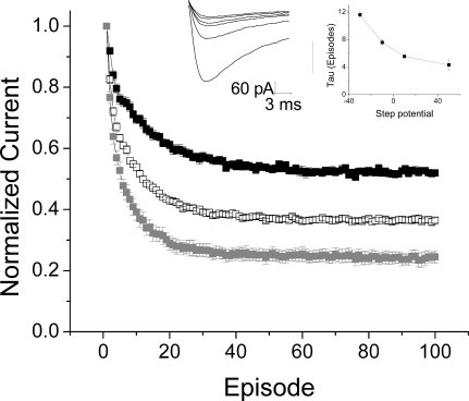
) (n = 8). Left inset, representative currents elicited by repetitive depolarizations from −110 to −10 mV with an interpulse interval of 100 ms in a cell recorded with 200 μM D575 included in the pipette. Right inset, τ values from exponential fits show voltage-dependence for D575 (■, n = 8).
Fig. 5.
Fig. 5.
Charged PAAs block CaV3.1 from the outside. A, mean current-voltage relationship ± S.E.M. produced in the absence (□) and presence of 20 μM D575 (▴, n = 5) or 20 μM qD888 (■, n = 5). Currents were normalized to peak current at −30 mV in the absence of drug. Top inset, fractional block at −10 mV produced by 20 μM D575 or qD888 is not significantly different from 20 μM verapamil; n = 3 for verapamil. Bottom inset, representative currents in the absence (top) and presence (bottom) of D575 (left) and qD888 (right). B, both 20 μM D575 and 20 μM qD888 produced use-dependent block. Mean peak currents ± S.E.M., normalized to the first pulse of the train and with mean control values subtracted; n = 3 for D575, n = 4 for qD888. Top inset, fractional block at the 30th pulse is not significantly different from block produced by 20 μM verapamil. Bottom inset, representative currents in the absence (left) and presence of D575 (middle) and qD888 (right).
Fig. 6.
Fig. 6.
MTSET can access the inner pore of CaV3.1 from the bath. A, a view of half of the inner pore of the T-type channel (the interface between domain III-S6 and domain IV-S6). Two of the four S6 helices (domains III and IV) are shown as green ribbons. Domain III-S5 is shown as a pink ribbon, and the domain III P-loop is shown in blue. Gln1828 and Leu1831, shown as space-filling images, are predicted to project toward the reader and into the inner pore. B, mean current-voltage relationship ± S.E.M. produced in the absence (□) and presence (■) of 2 mM MTSET in Q1828C channels, with 2 mM Ca2+ in the bath. Currents were normalized to peak current at −30 mV in the absence of MTSET; n = 3. Inset, sample traces recorded in the absence (top) and presence (bottom) of 2 mM MTSET.
Fig. 7.
Fig. 7.
An extracellular pathway for verapamil inside the inner pore of the T-type channel. Verapamil (shown in blue space-filling images) can be accommodated in the interface between domain III-S6, IV-S6 (shown by green ribbons), domain III-S5 (pink ribbon), and the domain III P-loop (blue ribbons). Some amino acid residues bordering this crevice are shown as space-filling images.
Fig. 8.
Fig. 8.
Insertion of a tryptophan in the back pathway reduces drug entry and exit. A, outside, mean peak currents ± S.E.M., recorded with 20 μM D575 in the bath, in WT (□, n = 3) and L1825W channels (■, n = 6). Currents were normalized to the first pulse of the train and mean control values were subtracted. Insets, representative currents elicited by repetitive depolarizations from −110 to −10 mV with an interpulse interval of 100 ms in a cell expressing L1825W, recorded with 20 μM D575 in the bath. τ Values from exponential fits show no change with voltage. B, inside: i, mean peak currents ± S.E.M., recorded with 200 μM D575 included in the pipette, in WT (□, n = 3) and L1825W channels (■, n = 4). Currents were normalized to the first pulse of the train, and mean control values were subtracted. Inset, representative currents elicited by repetitive depolarizations from −110 to −10 mV with an interpulse interval of 100 ms in a cell expressing L1825W, recorded with 200 μM D575 in the pipette. ii, τ values from exponential fits show voltage-dependence for D575 in both WT (□) and L1825W channels (■). iii, comparing the extent of block obtained with 200 μM D575 in WT (formula image
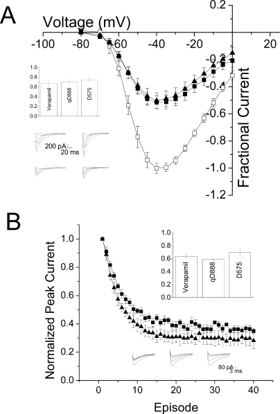
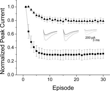
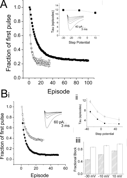
) and L1825W channels (□) shows some voltage-dependence in each case, with much greater block obtained in L1825W channels at the same drug concentration.

References

    1. Alpert LA, Fozzard HA, Hanck DA, Makielski JC. (1989) Is there a second external lidocaine binding site on mammalian cardiac cells? Am J Physiol 257:H79–H84 - PubMed
    1. Berjukov S, Aczel S, Beyer B, Kimball SD, Dichtl M, Hering S, Striessnig J. (1996) Extra- and intracellular action of quaternary devapamil on muscle L-type Ca2+-channels. Br J Pharmacol 119:1197–1202 - PMC - PubMed
    1. Creighton TE. (1993) Proteins: Structure and Molecular Properties, WH Freeman and Co., New York
    1. Cribbs LL. (2006) T-type Ca2+ channels in vascular smooth muscle: multiple functions. Cell Calcium 40:221–230 - PubMed
    1. Cheng RC, Tikhonov DB, Zhorov BS. (2009) Structural model for phenylalkylamine binding to L-type calcium channels. J Biol Chem 284:28332–28342 - PMC - PubMed

Publication types

MeSH terms

LinkOut - more resources