. 2010 Dec 28;107(52):22687-92.
doi: 10.1073/pnas.1012851108. Epub 2010 Dec 13.

CBP gene transfer increases BDNF levels and ameliorates learning and memory deficits in a mouse model of Alzheimer's disease

Affiliations

CBP gene transfer increases BDNF levels and ameliorates learning and memory deficits in a mouse model of Alzheimer's disease

Antonella Caccamo et al. Proc Natl Acad Sci U S A. .

Abstract

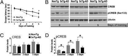
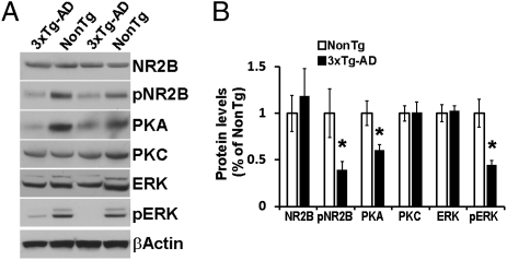
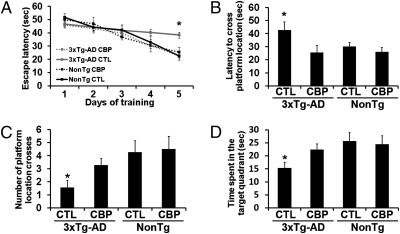
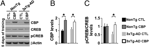
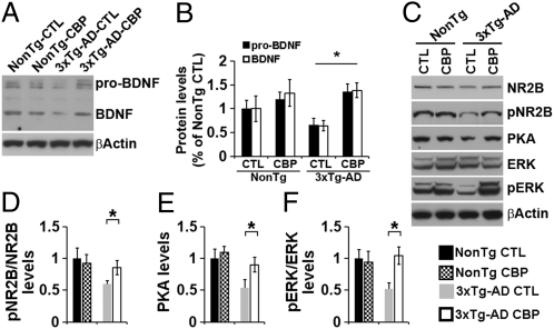
Cognitive dysfunction and memory loss are common features of Alzheimer's disease (AD). Abnormalities in the expression profile of immediate early genes that play a critical role in memory formation, such as the cAMP-response element binding protein (CREB), have been reported in the brains of AD patients. Here we show that amyloid-β (Aβ) accumulation, which plays a primary role in the cognitive deficits of AD, interferes with CREB activity. We further show that restoring CREB function via brain viral delivery of the CREB-binding protein (CBP) improves learning and memory deficits in an animal model of AD. Notably, such improvements occur without changes in Aβ and tau pathology, and instead are linked to an increased level of brain-derived neurotrophic factor. The resulting data suggest that Aβ-induced learning and memory deficits are mediated by alterations in CREB function, based on the finding that restoring CREB activity by directly modulating CBP levels in the brains of adult mice is sufficient to ameliorate learning and memory. Therefore, increasing CBP expression in adult brains may be a valid therapeutic approach not only for AD, but also for various brain disorders characterized by alterations in immediate early genes, further supporting the concept that viral vector delivery may be a viable therapeutic approach in neurodegenerative diseases.


Conflict of interest statement

The authors declare no conflict of interest.

Figures

Fig. 1.
Fig. 1.
Activity-dependent CREB activation is impaired in the 3xTg-AD mice. (A) Learning abilities of 6-mo-old 3xTg-AD and NonTg mice (n = 16/genotype) were evaluated in the spatial reference version of the MWM. At the end of the third day, eight mice per genotype were killed, and the remaining mice were trained for 2 additional days. On days 4 and 5 of training, the NonTg mice performed significantly better than the 3xTg-AD mice, as demonstrated by a shorter time to find the hidden platform. (B) Representative Western blots (probed with the indicated antibodies) of proteins extracted from the hippocampi of mice killed directly from their home cages or trained in an MWM for 3 d or 5 d. (C and D) Quantitative analyses of the blots show similar total CREB levels in the NonTg and 3xTg-AD mice at baseline and after neuronal stimulation. In contrast, at baseline, the pCREB levels were significantly reduced in the brains of 3xTg-AD mice compared with NonTg mice. Although the percentage increase in pCREB level did not differ significantly between the NonTg mice and the 3xTg-AD mice, the absolute pCREB levels were significantly lower in the 3xTg-AD mice at all of the time points analyzed. Protein levels are expressed as fold changes over sham-injected NonTg mice and represent mean ± SEM. *P < 0.05; **P < 0.01.
Fig. 2.
Fig. 2.
NMDA signaling is impaired in the 3xTg-AD mice. (A) Representative Western blots of proteins extracted from the hippocampi of 6-mo-old 3xTg-AD and NonTg mice (n = 8/genotype) and probed with the indicated antibodies. (B) Quantitative analysis of the blots shows that the levels of the NMDA receptor subunit NR2B phosphorylated at Tyr1472, PKA, and pERK were significantly decreased in the 3xTg-AD mice compared with the NonTg mice. Protein levels are expressed as fold changes over NonTg mice and represent mean ± SEM. *P < 0.05.
Fig. 3.
Fig. 3.
CBP gene transfer rescues learning and memory deficits in 3xTg-AD mice. CBP-expressing lentiviruses were injected into the dorsolateral ventricles of 3xTg-AD mice (n = 18) and NonTg mice (n = 17). In addition, 17 3xTg-AD mice and 16 NonTg mice received sham injections. Six mice per group were sacrificed after 3 d and 5 d of training. (A) All mice were evaluated in the spatial reference version of the MWM. The escape latency was significantly lower in the CBP-injected 3xTg-AD mice than in the sham-injected 3xTg-AD mice (P = 0.008). (B–D) Reference memory was significantly improved in the CBP-injected 3xTg-AD mice compared with the sham-injected 3xTg-AD mice in all probe trial measurements conducted. Data are presented as mean ± SEM. *P < 0.05.
Fig. 4.
Fig. 4.
CBP gene delivery restores pCREB levels after 5 d of training. (A) Representative Western blots of proteins extracted from the hippocampi of CBP- and sham-injected 3xTg-AD and NonTg mice (n = 6/group) and probed with the indicated antibodies. (B and C) Quantitative analysis of the blots shows significantly increased CBP levels in both the 3xTg-AD and NonTg mice receiving the virus. In contrast, pCREB levels were significantly increased in the CBP-injected compared with sham-injected 3xTg-AD mice, but not the NonTg mice. Data are presented as fold changes over sham-injected NonTg mice and represent mean ± SEM. *P < 0.05.
Fig. 5.
Fig. 5.
CBP gene delivery rescues BDNF levels. (A) Representative Western blots of proteins extracted from the hippocampi of CBP- and sham-injected 3xTg-AD and NonTg mice and probed with an anti-BDNF antibody. (B) Quantitative analysis of the blots shows that in the hippocampi of the 3xTg-AD mice, CBP gene delivery restored the levels of pro-BDNF and BDNF to NonTg levels. (C) Representative Western blots of proteins extracted from the hippocampi of 6-mo-old sham- and CBP-injected 3xTg-AD and NonTg mice (n = 6/genotype) after 5 d of training and probed with the indicated antibodies. (D–F) Quantitative analysis of the blots shows that after 5 d of training, the pNR2B, PKA, and pERK levels were significantly increased in the CBP-injected 3xTg-AD mice compared with sham-injected 3xTg-AD mice. In contrast, no differences were found between the sham- and CBP-injected NonTg mice. Data are presented as fold changes over sham-injected NonTg mice and represent mean ± SEM. *P < 0.05.

References

    1. Querfurth HW, LaFerla FM. Alzheimer's disease. N Engl J Med. 2010;362:329–344. - PubMed
    1. Crews L, Masliah E. Molecular mechanisms of neurodegeneration in Alzheimer's disease. Hum Mol Genet. 2010;19(R1):R12–R20. - PMC - PubMed
    1. Snyder EM, et al. Regulation of NMDA receptor trafficking by amyloid-beta. Nat Neurosci. 2005;8:1051–1058. - PubMed
    1. Palop JJ, Mucke L. Amyloid-beta–induced neuronal dysfunction in Alzheimer's disease: From synapses toward neural networks. Nat Neurosci. 2010;13:812–818. - PMC - PubMed
    1. Dickey CA, et al. Selectively reduced expression of synaptic plasticity-related genes in amyloid precursor protein + presenilin-1 transgenic mice. J Neurosci. 2003;23:5219–5226. - PMC - PubMed

Publication types

MeSH terms

Substances